Participation in ultramarathon races and knowledge of these athletes continues to increase as the sport becomes more popular. Physicians and athletes need to better understand the impact of the unique aspects of ultramarathon races, such as race environment (temperature, humidity, and altitude), race distance, race stages, nutritional requirements and equipment, on athlete injuries and illness. Proper treatment of injuries and illnesses during an ultramarathon race is important for avoiding long-term medical issues. In this article, the evaluation and treatment of common musculoskeletal injuries and medical illnesses in ultramarathon runners are reviewed.
Key points
- •
In ultramarathoners, most musculoskeletal and skin-related issues are minor and can be treated successfully during the course of a race.
- •
Common medical illnesses, including hyperthermia and exercise-associated hyponatremia, require prompt assessment.
- •
Multistage ultramarathoners are more likely to experience hypernatremia than hyponatremia.
- •
Continued research should focus on preventative and optimal treatment strategies in hopes of preventing long-term complications in this unique athletic population.
Introduction
Ultramarathon races represent any foot race longer than 42 km; an estimated 70,000 runners participate yearly in running races throughout the world. Most races are single-stage point-to-point continuous races occurring over a specific period (ie, 1 to 2 days). Multistaged races are point-to-point races that occur over 3 to 7 days. Ultramarathons typically occur in more extreme environments with variations in terrain (mountains, snow, sand dunes, river crossings, slot canyons), temperature, and humidity. Ultramarathon runners require different equipment depending on the length and environment of the race. In multistage races, runners must be prepared to carry all their gear (eg, food, water, protective clothing) throughout the race.
Our knowledge of common injuries and illnesses in ultramarathon runners continues to increase. In the 1990s, studies focused on musculoskeletal injuries, noting that most running-related injuries were caused by Achilles tendinopathy (2%–18%) and patellofemoral pain (7%–15%). In 2011, Krabak and colleagues’ prospective study of multistage ultramarathon runners suggested that 95% of injuries are minor and are caused by skin-related disorders (74.3%), musculoskeletal injuries (18.2%), and medical illnesses (7.5%) ( Table 1 ). Other studies have focused on often asymptomatic but potentially injurious diseases, like exercise-associated hyponatremia (EAH) and acute kidney injury (AKI), reporting an EAH incidence of 8% to 50% and AKI in more than 50% of the studied athletes. Little is known about cardiovascular events during ultramarathon races.
| Diagnosis | Marathon, n (%) | Multistage Ultramarathon | |
|---|---|---|---|
| Major, n (%) a | Minor, n (%) a | ||
| Medical illnesses | |||
| Exercise-associated collapse b | 863 (59.4) | 35 (56.5) | 43 (3.9) |
| Altitude sickness | — | 0 | 11 (1.0) |
| Serious medical diagnosis c | 2 (0.14) | 1 (1.6) | 1 (0.1) |
| Other medical diagnosis d | 7 (0.48) | 0 | 27 (2.4) |
| Musculoskeletal injuries | |||
| Bursitis | — | 1 (1.6) | 11 (1.0) |
| Sprain | 19 (1.3) | 2 (3.2) | 25 (2.3) |
| Strain | 207 (14.3) | 1 (1.6) | 27 (2.4) |
| Tendonitis | — | 7 (11.3) | 115 (10.3) |
| Other e | 4 (0.28) | 3 (4.8) | 29 (2.6) |
| Skin disorders | |||
| Abrasion | 27 (1.9) | 0 | 43 (3.9) |
| Blister | 289 (19.9) | 10 (16.2) | 642 (57.8) |
| Cellulitis | — | 1 (1.6) | 8 (0.7) |
| Hematoma (subungual) | — | 1 (1.6) | 106 (9.5) |
| Other f | — | 00 | 23 (2.1) |
a Major, unable to continue in race; minor, able to continue in race.
b Hyperthermia, normothermia, hypothermia.
c Hyponatremia, hematuria, renal stone.
d Blurry vision, conjunctivitis, diarrhea, dyspepsia, epistaxis, hematochezia, insect bite, neuropathy, pharyngitis, upper respiratory infections.
e Fracture, metatarsalgia, contusion, costochondritis, laceration, splinter.
With the increase in ultramarathons come inherent challenges relating to the unique environments, training demands, nutritional preparation, and equipment. These challenges provide education and research opportunities for both physicians and athletes. Ill-prepared athletes and physicians place the athlete at risk for injury and illness. By better understanding the ultramarathon athlete, the sports medicine physician can provide optimal care and it is hoped limit morbidity and mortality. In this article, the understanding of strategies for managing commonly encountered musculoskeletal injuries and medical illnesses in ultramarathon runners is reviewed.
Musculoskeletal injury
Injuries to the musculoskeletal system are common in running sports. Reported musculoskeletal injury incidence varies depending on the methodology of the study. Musculoskeletal injury rates range from 2% to 18% in continuous single-stage ultramarathons and 19% to 22% in multistage, multiday ultramarathons. In multistage, multiday ultramarathons, musculoskeletal injuries accounted for 18% of the minor encounters (able to continue racing) and 22% of the major injury encounters (unable to continue racing) and are most likely to occur during stages 3 or 4 of a 7-stage race, highlighting the potential cumulative effect on the musculoskeletal system of running long distances. Although general muscle soreness affects most ultramarathoners, true musculoskeletal injuries, whether minor or major, may decrease performance and result in decreased training or medical withdrawal from a race.
Lower extremity injuries predominate, with the knee and ankle being most affected. Evaluation and treatment of these injuries may differ in an acute setting during or immediately after a race compared with the subacute or chronic setting in a standard medical clinic setting. For the purposes of this article, the focus is the acute race or outpatient setting:
Achilles Tendinopathy/Tendonitis
Studies suggest a prevalence of 2.0% to 18.5% and incidence of 10.8%. The mechanism of injury is usually repeated or strained plantar flexion. Runners present for evaluation complaining of posterior heel or ankle pain. On physical examination, swelling or fullness may be visible in the affected Achilles region, although this is not always present. There is tenderness to palpation or squeezing of the tendon, most commonly at the midportion of the tendon, where there is a watershed vascular region of relatively low blood supply. Alternatively, the focus of tenderness may be at the insertion of the tendon at the posterior calcaneous, indicating that an enthesopathy or bursitis may be contributing to the pain. The runners may have pain with passive, manual stretch of the Achilles tendon. They usually have pain and possibly weakness with repeated single-leg toe raises and may be unable to perform full excursion for 5 to 10 repetitions. This method of testing strength is preferable to having them plantarflex against manual resistance, because their Achilles strength is greater than any examiner’s upper extremity–provided resistance, meaning a manual test would miss all but the most severe cases. A Thompson test (passive manual calf squeeze showing plantar flexion if Achilles is intact) should be performed to rule out a significant tendon tear. Treatments in the acute setting include icing or cold compress for 15 to 20 minutes (repeated several times during the evening while not running), gentle stretching of the tendon, and providing an overnight dorsiflexion splint or bandage wrap. If elastic, stretchy tape is available, application of plantarflexion assist elastic tape can help during ambulation or continued running but should be removed at night in favor of dorsiflexion splinting. Topical antiinflammatory creams can be used at any time if the overlying skin is intact. Oral analgesics like acetaminophen can be used to help decrease pain. Current guidelines recommend that oral nonsteroidal antiinflammatory (NSAID) medications should be used only at the end of the day/race section when the runner can be adequately hydrated to prevent AKI associated with NSAID use. In the subacute setting, sports medicine clinical treatment strategies may include rest, physical therapy with a focus on eccentric exercises, peritendon steroid injections, intratendon autologous blood product injections, or referral to surgery for recalcitrant cases.
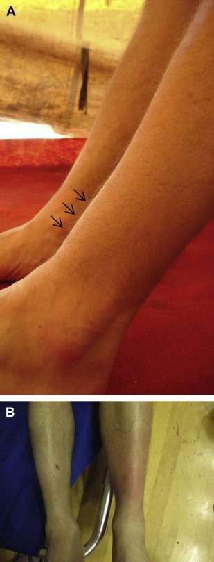
Anterior Tibialis Tendinopathy/Tendonitis
The mechanism of injury is usually repeated or strained dorsiflexion (particularly on a course that has prolonged, steep hills/inclines). Runners present for evaluation complaining of anterior ankle pain. On physical examination, swelling or fullness may be visible in the affected anterior tibialis tendon ( Fig. 1 A), although this is not always present. Occasionally, there is associated erythema in the anterior ankle as well (see Fig. 1 B), and this must be closely followed to rule out a concurrent infection/cellulitis. They are tender to palpation of the tendon. They may have pain with passive, manual stretch of the anterior tibialis tendon, which is best elicited with combined passive ankle plantar flexion and passive great toe plantarflexion. The runners usually have pain and possibly weakness with repeated resisted ankle dorsiflexion, because they should be able to support the examiner’s application of upper body weight (not simply the examiner’s arm/hand strength) against dorsiflexed ankles. Treatments in the acute setting include icing or cold compress for 15 to 20 minutes (repeated several times during the evening while not running), and gentle stretching of the tendon. Topical antiinflammatory creams can be used at any time if the overlying skin is intact. In multistage races, anecdotal cases of using an abundance of topical cream over the tendon with a bandage or occlusive dressing overnight has worked well, but it should be used with care and attention to possible skin reaction/allergy. Oral medications (analgesics and NSAIDs) follow the same guidelines as in the section on Achilles tendonitis. In the subacute setting, sports medicine clinical treatment strategies may include rest, physical therapy, peritendon steroid injections, intratendon autologous blood product injections, or referral to surgery for recalcitrant cases.
Plantar Fasciitis
Studies have suggested an incidence of 10.6%. The mechanism of injury is repeated impact/shock absorption without proper arch support (either lack of support or too much support/pressure) or with higher-impact activities, even if arch support is correct for the corresponding anatomy and running style. Runners present for evaluation complaining of pain in the sole of the foot, frequently midarch, but it can be just proximal or distal to that. On physical examination, they are usually tender to palpation at the midarch or proximally along the plantar fascia toward the origin at the calcaneous. Pain may be reproduced with passive manual dorsiflexion, particularly if combined with toe extension. The runners may also have pain with resisted toe flexion in combination with ankle plantarflexed positioning. Treatment includes ice to the area, particularly ice massage by rolling a frozen or very cold water bottle underneath the arch after the day’s running is complete. Passive manual stretch of the plantar fascia and dorsiflexion splinting at night can also help improve symptoms. The same medication guidelines given earlier for Achilles tendonitis hold true. In the subacute setting, strengthening intrinsic muscle of the foot, running gait evaluation/recommendations, and trials of varying levels of arch support/shoe style can help treat symptoms and prevent recurrence. Rest, physical therapy, corticosteroid injections, autologous blood product injections, and referral to surgery may also be used in an outpatient treatment setting.
Patellofemoral Syndrome
The prevalence in ultramarathon is 7.4% to 15.6%. Incidence of knee issues is 24%, although it is not clear how many of these are patellofemoral versus other knee issues (eg, meniscus tear, osteoarthritis). This injury tends to occur more often in the chronic setting but can occur as acute pain in ultradistance races. Mechanism of injury can be underlying structural anomaly of shallow femoral groove or lateralization of the patella versus inadequate quadriceps and core muscle strength for the demand distance running. Runners present with anterior knee pain. There are usually no symptoms of frank buckling or locking, but the injury can be associated with grinding sensation or crepitus. On physical examination, there is tenderness on the patellar facets, usually more on the medial aspect. There may also be tenderness along the patellar tendon, indicating a component of patellar tendonitis/tendinopathy as well. Pain may be reproduced with single-leg or double-leg squat or resisted knee extension. Acute treatment includes ice to the area and oral medications, as described earlier, taking care not to ice if the runner plans to continue running that day. A patellar tracking knee brace or band strap may help decrease pain with ambulation and running. Athletic tape can be used to fabricate a patellar band strap if none is readily available. If practitioners are trained in using kinesio tape for patellar tracking, that may also be helpful. In the subacute setting, physical therapy with attention to quadriceps and core strengthening can be used. Rest, steroid injections to the knee, viscosupplement injections if arthritis is associated, or referral to surgery for release of the lateral retinaculum or other interventions may also be used as treatment strategies.
Ankle Sprain
The incidence in ultramarathons is 10.8%. The mechanism of injury is usually an inversion rolling of the ankle on uneven terrain, causing strain or tear of any of the anterolateral ligaments of the ankle. The runner presents with anterior or lateral ankle pain. There may be swelling or ecchymosis, and the athlete may or may not be able to fully bear weight. On physical examination, the athlete has tenderness to palpation over the affected ligaments. Tenderness over the bone/lateral maleolus may indicate a fracture. If able to bear weight, they may have increased pain or instability when standing on the toes with the ankles plantarflexed. An anterior drawer test may show laxity compared with the contralateral side. Initial treatment consists of rest, ice, compression, and elevation. If any instability or ecchymosis is noted, or if there is a high suspicion of fracture (based on Ottawa ankle rules), they should be removed from competition. Oral medications can be used as described in the section on Achilles tendonitis. If fracture is suspected, the patient must remain non–weight bearing until transported to a facility with radiograph services for further diagnostic evaluation. Medial-lateral ankle brace support or a rocker bottom boot should be used in cases of instability in which fracture is not suspected. A lace-up ankle brace or athletic taping of the ankle can be used for those who have no instability and are able to walk without a limp to allow them to continue if competition if desired.
Muscle Strain
The most common muscle strains involve the calf (incidence 13.1%) and hamstring (incidence 11.8%). Mechanism of injury is usually an eccentric contraction or quick burst movement, which stresses the muscle to the point of injury without tearing a significant number of muscle fibers (although a small tear is difficult to differentiate from a strain). Most frequently involved muscles are those that cross 2 joints, like the hamstrings (crossing both hip and knee joints) and the gastrocnemius (crossing both knee and ankle joints) Runners present with acute pain in the affected muscle. On physical examination, they may have pain with passive stretch or active contraction of the muscle. Initial treatment consists of rest, ice (after the day’s competition is finished), compression, and elevation, as noted earlier. Oral medications can be used as described in the section on Achilles tendonitis. A runner may be able to continue in competition with a mild strain, but anything that causes limping or altered gait should be cause to consider removing the athlete from competition for risk of further injury. If a full tear of a muscle is suspected, then they should be removed from competition immediately and referred for further evaluation at a hospital or sports medicine clinic. In the subacute setting, if relative rest for 1 to 2 weeks along with oral antiinflammatory medication and the other treatments noted earlier do not resolve the issue, then, physical therapy or advanced imaging studies may be warranted.
Iliotibial Band Problem
The incidence is 15.8%. Mechanism of injury is usually an overuse, more chronic presentation in runners but can be acute overuse in ultradistance running. The prevailing theory links this entity to impingement of the distal iliotibial band (ITB) at the lateral femoral condyle during the eccentric contraction just after heel strike. Runners present with lateral knee pain at the lateral femoral condyle or just proximally or distally. They may have exacerbation of pain with compression of the band/tendon over the femoral condyle (Noble compression test) or with stretch of the ITB (Ober test). Treatment during competition consists of stretching the ITB and using topical antiinflammatory cream locally (if overlying skin is intact). Oral medications can be used, as described earlier. Icing should be used at the end of the day’s competition, as described earlier. Cross-fiber friction massage can be used in the acute setting. In the subacute setting, rest, physical therapy, deep massage with foam roller, gait analysis, flexibility training, and core strengthening can be used for treatment. Other injections (eg, steroid, autologous blood products) may be considered if conservative treatment options fail.
Back Injuries
The incidence is 12.4%. Mechanism of injury can be varied, from acute muscle strain, spasm or disk injury to chronic degenerative change in the lumbar spine. Runners presenting with acute axial back pain without trauma may be treated as a strain (rest, ice oral medications) or if believed to have palpable spasm, then application of heat and gentle stretching are appropriate treatments. If the presentation includes pain radiating into the thigh or lower leg, or if lower extremity numbness/tingling is associated, then, they may have an acute lumbar radiculopathy and should be removed from competition, given oral antiinflammatory medication, and referred for nonemergent evaluation at a hospital or clinic. Any presentation of back pain with notable lower extremity weakness, numbness/tingling in the groin/genitals/rectal area, or loss of control of bladder or bowel function should be treated as a spinal emergency and the patient should be transported to a hospital for immediate evaluation and treatment.
Related posts:

Stay updated, free articles. Join our Telegram channel

Full access? Get Clinical Tree


