CHAPTER 1 Elbow Arthroscopy: Positioning, Setup, Anatomy, and Portals
Elbow arthroscopy is a technically demanding procedure, and extensive hands-on training and supervised experience are needed to acquire proficiency. When performed with appropriate judgment and technique, elbow arthroscopy is an excellent tool for the correction of many lesions of the elbow joint with minimal risk.1 However, it poses greater technical challenges and neurologic risks than knee or shoulder arthroscopy. Arthroscopy of the elbow joint is perhaps the most hazardous in terms of its potential for causing injury to nearby nerves and vessels because of the complex relationship of these structures to the joint (Fig. 1-1).2 Because of the surrounding neurovascular structures, familiarity with the normal elbow anatomy and portals can decrease the risk of damage to important structures.3 Elbow arthroscopy provides an opportunity for diagnostic and therapeutic intervention with little morbidity.
Burman4 first described elbow arthroscopy in 1931, but he stated that the elbow is “… unsuitable for examination since the joint is so narrow for the relatively large needle.” In 1932, he revised his opinion based on the arthroscopic examination of 10 cadaveric elbows.5 After Burman’s studies were published, a small number of reports appeared in the German and Japanese literature, but it was not until the middle to late 1980s that reports began to appear in the American literature.6,7
In 1985, Andrews and Carson7 described the patient-supine technique and the use of various portals for elbow arthroscopy. In 1989, Poehling and colleagues8 described the patient-prone position for elbow arthroscopy. Since then, the techniques and indications for elbow arthroscopy have expanded, and there have been many more reports describing variations in operative technique.
ANATOMY
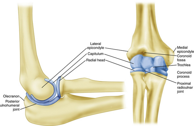
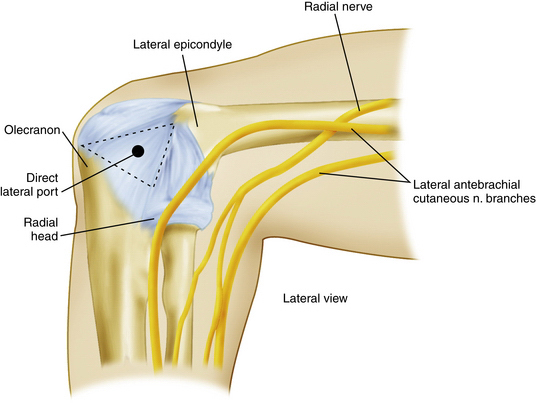
A clear understanding of the anatomy of the elbow is important before proceeding with arthroscopy. Important bony anatomic landmarks that should be palpated include the lateral and medial epicondyles, olecranon process, and radial head (Fig. 1-2). On the lateral side, the lateral epicondyle, olecranon process, and radial head form a triangle. Located in the center of this triangle is a soft spot called the anconeus triangle. It often is used to inflate the joint with fluid before introducing instruments or cannulas, and it can be the landmark for a direct lateral portal (Fig. 1-3). Posteriorly, important structures include the triceps muscle, tendon, and tip of the olecranon.
Sensory nerves around the elbow include the medial brachial cutaneous, the medial antebrachial cutaneous, the lateral antebrachial cutaneous, and the posterior antebrachial cutaneous nerves.9 The medial brachial cutaneous nerve penetrates the deep fascia midway down the arm on the medial side, and it supplies skin sensation to the posteromedial aspect of the arm to the level of the olecranon. The medial antebrachial cutaneous nerve supplies sensation to the medial side of the elbow and forearm. The lateral antebrachial cutaneous nerve is a branch of the musculocutaneous nerve, which exits between the biceps and brachialis muscles laterally to supply sensation to the elbow and lateral aspect of the forearm. The posterior antebrachial cutaneous nerve branches from the radial nerve and courses down the lateral aspect of the arm to supply sensation to the posterolateral elbow and posterior forearm.6
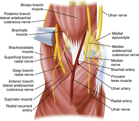
The main neurovascular structures about the elbow are the median nerve, radial nerve, ulnar nerve, and brachial artery.9 The radial nerve spirals around the posterior humeral shaft, penetrates the lateral intermuscular septum, and descends anteriorly to the lateral epicondyle between the brachioradialis and brachialis muscles. The radial nerve then branches to form the superficial radial nerve, which supplies sensation to the dorsoradial wrist and posterior surface of the radial three and one-half digits, and the posterior interosseous nerve, which provides motor branches to the wrists, thumb, and finger extensors. The ulnar nerve penetrates the medial intermuscular septum in the distal one third of the arm, courses posteriorly to the median epicondyle, and then descends distally between the flexor carpi ulnaris and flexor digitorum superficialis muscles. The brachial artery courses just medial to the biceps tendon in the antecubital fossa and then descends to the level of the radial head, where it bifurcates into the radial and ulnar arteries (see Fig. 1-1).6
PATIENT EVALUATION
History
The differential diagnosis for symptoms of the anterior elbow includes distal biceps tendon rupture, which can be partial or complete; anterior capsular strain; and brachioradialis muscle strain.10 Symptoms in the posterior compartment can reflect valgus extension overload syndrome, posterior impingement, osteochondral loose bodies, triceps tendonitis, triceps tendon avulsion, or olecranon bursitis.11 Deep, aching pain in the posterior region of the elbow may indicate an olecranon stress fracture.12
A careful neurovascular history is also important as ulnar nerve paresthesias can be the result of cubital tunnel syndrome, a subluxing ulnar nerve, or a traction injury from valgus instability.6
Throwing athletes are a unique patient population, and it is important to gather information about prior injury and any changes in the throwing mechanism or rehabilitation regimen.10 A patient whose symptoms are related to throwing and are located medially may have an injury to the medial collateral ligament. Throwing athletes who report lost velocity and control or an inability “to let the ball go” may have pain posterior on forced extension, which may signify posterior olecranon impingement caused by a medial collateral ligament injury. The typical patient is a baseball pitcher in his mid-20s who has posterior elbow pain during the acceleration and follow-through phases of pitching and complains of the inability to fully extend the elbow.6 Young throwing athletes (<18 years) with OCD lesions often report progressive lateral elbow pain during late acceleration and follow-through phases, with loss of extension and episodes of locking.11
Physical Examination
A careful physical examination of all three compartments of the elbow is critical to determine the correct diagnosis. Each compartment should be examined individually to fully evaluate the elbow. The physical examination starts with careful inspection of the skin and soft tissues to make sure there are no scars, swelling, ecchymosis, soft tissue masses, or bony abnormalities. The alignment of the elbow should be inspected, noticing any significant varus or valgus deformities. Range of motion of the elbow in flexion, extension, supination, and pronation should be observed and compared with the contralateral side. Those with posteromedial impingement or valgus extension overload may reveal a flexion contracture and pain over the posteromedial olecranon tip.6
The examiner should test for valgus instability with the elbow flexed to 30 degrees to relax the anterior capsule and free the olecranon from its bony articulation in the olecranon fossa. A valgus stress is then applied with the elbow in full supination. Discomfort along the medial aspect of the elbow can indicate ulnar collateral ligament injury. Valgus laxity, however, is often difficult to discern, particularly if there is tearing of the undersurface of the ulnar collateral ligament.13 Comparison with the contralateral elbow can help differentiate physiologic laxity from pathologic instability.14
The triceps muscle insertion and the posterolateral and posteromedial joint areas are palpated to assess for tenderness, bone spurs, and posterior impingement lesions. The so-called clunk test is performed to demonstrate posterior olecranon impingement.15 The upper arm is grasped and stabilized as the elbow is brought into full extension. Reproduction of pain at the posteromedial aspect of the joint suggests compression of the olecranon into the fossa and indicates valgus extension overload.
The lateral epicondyle and extensor origin are palpated to assess for lateral epicondylitis. The radiocapitellar joint is palpated while the forearm is pronated and supinated to elicit crepitus or catching, which can be caused by chondromalacial lesions or impingement from a lateral synovial fringe.16 The soft spot is also inspected to determine whether there is synovitis or an effusion in the elbow joint.6
Stability can be assessed with O’Driscoll’s posterolateral rotatory instability test.17 The test is best done under general anesthesia because of the patient’s apprehension while awake, which may give a false-negative result. However, it can be done with the patient awake with the extremity over the patient’s head and the shoulder in full external rotation. During the test, a valgus, supination, and axial compression load is applied to the elbow, which is flexed approximately 20 to 30 degrees. With the elbow in extension, subluxation or dislocation of the radius and of the proximal ulna creates a posterior prominence and sulcus sign. When the elbow is flexed, radiohumeral and ulnohumeral joints are visibly or palpably reduced.6 Details of this technique can be found in Chapter 11.
Diagnostic Imaging
Routine diagnostic radiographs include an anteroposterior view with the elbow in full extension and a lateral view with the joint in 90 degrees of flexion. An axial view can be obtained to outline the olecranon and its medial and lateral articulations. This is the best view for identifying and assessing a posteromedial osteophyte. When there is a history of trauma, an oblique view should be done, and careful attention should be paid to the radial head and the coronoid process for subtle fracture lines. Radiographs should be reviewed for more obvious anterior or posterior elbow dislocations, along with more subtle degenerative changes, osteophytes, and loose bodies. However, plain radiographs are not always able to demonstrate all loose bodies.18
A gravity stress test radiograph can be used to detect valgus laxity of the elbow.19 The patient is placed in a supine position, and the shoulder is abducted and brought to maximum external rotation so that the elbow is parallel to the floor. If there is an injury to the ligament or bony attachment, increased joint space can be seen on radiographs.6
Magnetic resonance imaging (MRI) and computed tomography (CT) arthrography (CTA) have are accurate in diagnosing a complete tear of the ulnar collateral ligament.13 However, in early studies, CTA was more sensitive in detecting a partial undersurface tear of the ulnar collateral ligament.20 This was described as a T-sign lesion by Timmerman and Andrews, who said that it represents “…dye leaking around the detachment of the deep portion of the ulnar collateral ligament from its bony insertion, but remaining within the intact superficial layer, ulnar collateral ligament, and capsule.”13
MRI is useful for evaluating osteochondral lesions in the radiocapitellar joint21,22 and for demonstrating early vascular changes that are not yet apparent on plain radiographs, and it can be used to assess the extent of the lesion and displacement of fragments.6 MRI is also helpful for evaluating the soft tissue structures of the elbow, including the tendinous insertions of the flexor and extensor musculature to help in diagnosing medial and lateral epicondylitis, the triceps insertion and associated musculature to evaluate for triceps tendonitis, and the medial and lateral collateral ligaments for possible tears. However, MRI may not demonstrate subtle undersurface tears of the ulnar collateral ligament. Magnetic resonance arthrography with saline contrast or gadolinium can increase the sensitivity for detecting undersurface tears of the ulnar collateral ligament and has become the test of choice to detect these tears.13
TREATMENT
Indications and Contraindications
In 1992, O’Driscoll and Morrey described the early indication for elbow arthroscopy, which was pain or symptoms that were substantial enough to interfere with work, daily activities, sports, or sleep and that did not resolve after conservative treatment.23 In this early study, they analyzed the results of 71 elbow arthroscopies as the indications for such a procedure were evolving. Not surprising, the best early results were seen for arthroscopic removal of loose bodies, assessment of undiagnosed snapping, idiopathic flexion contractures, local débridement of damaged articular surfaces, and synovectomy. They found that the patients least likely to benefit were the ones in whom there was a disparity between objective and subjective findings.23
Since then, the indications for elbow arthroscopy have evolved. In 1994, Poehling further refined the indications, which included its use for the diagnosis of intra-articular lesions of the elbow, the removal of loose and foreign bodies, irrigation of the joint, débridement of an infected joint, excision of osteophytes, synovectomy, capsular release, excision of the radial head, and treatment of acute fractures of the elbow.1
Several investigators have since reported the usefulness of elbow arthroscopy for the removal of loose bodies,6,7,18,23-25 and this continues to be the primary indication for elbow arthroscopy. Several pathologic processes may initiate the formation of a loose body, including trauma and synovial chondromatosis. Regardless of the cause, the patients usually present with swelling, locking, pain, and loss of motion, all of which can be improved with the removal of loose bodies. These loose bodies can be found in the anterior and posterior compartments and in the posterior medial gutter, and removing them can be a technically demanding procedure. Further details and helpful hints can be found in Chapter 2.
Elbow arthroscopy can be an effective tool if the diagnosis of an infection is made or suspected. It is a less invasive way to enter into the joint with minimal trauma to confirm the diagnosis of an infection, irrigate the joint, débride infected tissue, and assess the condition of the underlying bone, cartilage, and synovial tissue.1
The presence of osteophytes, or osseous spurs, is another condition that lends itself to arthroscopic management and removal.23–26 A true lateral radiograph of the elbow is useful for the identification of osteophytes that may limit full extension of the elbow with impingement of the posterior olecranon spur in the olecranon fossa.1,23 An axial view may also show a posteromedial osteophyte,6 which can be easily removed arthroscopically.
The term valgus extension overload was coined27 to describe the findings that can be identified in baseball pitchers and other overhead athletes. The tremendous repetitive valgus forces generated during the acceleration and follow-through phases of pitching, as the elbow goes into extension, can result in osteochondral changes in the olecranon and distal humerus. A significant osteophyte forms on the posteromedial aspect of the olecranon fossa with continued pitching or overhead activities, creating an area of chondromalacia.6 The use of elbow arthroscopy in the throwing athlete, including evaluation of the medial collateral ligament, is addressed in Chapter 12. The inability to reliably visualize the anterior bundle of the medial collateral ligament with the arthroscope limits the value of the arthroscope when assessing medial collateral ligament injuries.28,29 The management of posterolateral instability is discussed in Chapter 11.
Chronic synovitis caused by inflammatory arthritis that does not respond to nonoperative management and when there is minimal joint destruction can be an indication for elbow arthroscopy. Synovectomy can provide considerable relief of pain.23,30,31 Diagnostic elbow arthroscopy can also be used for synovial biopsy to establish the diagnosis of rheumatoid arthritis or other inflammatory arthritides or a monoarticular or polyarticular arthritis of unknown origin.23 The elbow joint is affected in approximately 20% to 50% of patients with rheumatoid arthritis, and 50% of these patients develop pain and associated loss of motion.23,32 Lee and Morrey achieved a 93% rate of good or excellent results in a short-term follow-up assessment of 14 arthroscopic synovectomies in 11 patients. However, only 57% of their patients maintained good or excellent results at an average of 42 months after surgery.31 When performing elbow arthroscopy and synovectomy for rheumatoid arthritis and other inflammatory arthritides, it is important to set realistic expectations for the patient because the symptoms can recur.
OCD of the capitellum is characterized by pain, swelling, and limitation of motion, and it usually occurs during adolescence or young adulthood in a throwing athlete or gymnast.33 The underlying cause of this lesion is most likely repetitive microtrauma to a vulnerable epiphysis with a precarious blood supply.33 The lesion may progress to joint incongruity, loose body formation, and a locked elbow with chronic pain, all of which are indications for elbow arthroscopy when conservative measures fail. The procedure involves the arthroscopic removal of osteophytes, excision of loose or detached cartilage, and curettement and drilling of the base of the lesion.34
Panner’s disease is an osteochondrosis of the entire capitellum in children and adolescents, and it may represent an early stage of OCD.6 Reconstitution of the capitellum usually occurs with rest and without late sequelae or limitations.33 More information about OCD can be found in Chapter 7.
The indications for arthroscopic débridement of the elbow for degenerative osteoarthritis are similar to those described for loose body removal, valgus extension overload, and OCD. Pain associated with swelling and mechanical symptoms of catching and locking respond well to arthroscopic débridement.24

Stay updated, free articles. Join our Telegram channel

Full access? Get Clinical Tree


