Diagnosis and Management of Extremity Vascular Injuries


Diagnosis and Management of Extremity Vascular Injuries


C. Kristian Enestvedt

David Cho

Donald D. Trunkey


INTRODUCTION

Extremity vascular injuries continue to pose clinical challenges to trauma and vascular surgeons. More than three fourths of all vascular trauma occurs in the extremity.1,2,3,4,5 Although most clinically significant peripheral vascular injuries result from penetrating trauma, the contribution to this cohort from blunt trauma may be higher depending on the injury mechanism and setting.2,4,5,6,7,8,9,10,11,12,13,14 The accumulated experience with vascular trauma has seen considerable contributions from surgeons in combat settings, where most injuries result from penetrating trauma. Historically, many arterial injuries were managed with simple ligation followed by amputation. Current management yields high rates of successful vascular repair and significantly improved limb salvage rates compared to vessel ligation. In addition, the development of endovascular techniques will impact the future of surgical management for vascular trauma.


Patient Evaluation

The evaluation of trauma victims with vascular injury should follow the basic protocols of Advanced Trauma Life Support (ATLS). A thorough, focused vascular examination is paramount when an arterial injury is suspected. Palpation of the arterial pulse at all levels in the affected limb(s) should begin distally and move proximally, with note made of the quality, strength, and rate at each level. Throughout the examination, the injured limb should be compared to the contralateral limb. Occasionally, hard signs of arterial disruption will be encountered such as active arterial hemorrhage, expanding hematoma, limb ischemia, thrill, bruit, or absent pulses (see Table 1). Of these, pulse deficit is reportedly the most common.15 Although compression techniques-including placement
of tourniquets-can be used as temporizing measures for active arterial hemorrhage in the trauma bay, further management should be performed in the operating room. Radiographic or ultrasonographic assessment of a limb with an absent pulse, however, is obligatory if other issues do not require immediate attention in the multiply injured patient.


TABLE 1 HARD AND SOFT SIGNS OF VASCULAR INJURY


Hard signs


Arterial bleeding


Ongoing hemorrhage with shock


Pulsatile hematoma


Limb ischemia


Bruit or thrill


Loss or absence of distal pulses


Soft signs


Nonexpanding hematoma


Injury to nerve associated with vessel


Wound or injury tract <1 cm from major vessel


Decreased ABI


Knee dislocation


History of massive bleeding now stopped


ABI, Ankle Brachial Index.


Evaluation of the affected extremity with a handheld Doppler ultrasound should be performed routinely in all patients with nonpalpable pulses. It is important to make note of the phase pattern of the waveforms. Absent Doppler signals warrant further imaging—either with angiography, duplex ultrasonography, or computed tomography with angiogram (CTA). Every Doppler examination, whether normal or abnormal, should be followed by the Ankle Brachial Index (ABI) measurement. An ABI >1.0 is considered normal. Any measurement <0.9 warrants further evaluation, usually with angiography. Again, it is important to compare findings between limbs.

Combined with ABI, the physical examination (PE) has proved to be an accurate measure of arterial injury.8,15,16,17,18,19 A high negative predictive value has been well documented when the PE is used as a screening test in the trauma setting, with fewer than 1.5% of occult vascular injuries ultimately requiring operative intervention.16 The robust sensitivity of screening with PE and ABI has been validated by multiple authors for both penetrating and blunt injury. Gonzalez et al.15 examined the utility of PE for the evaluation of penetrating extremity trauma in 406 patients with 489 vascular injuries. The authors demonstrated a sensitivity of 92% and a specificity of 95%. Their screening protocol included PE for hard signs of vascular injury, assessment of injury trajectory, and Doppler ultrasound. Those patients without hard signs were observed and those with hard signs were either taken directly to the operating room or for angiography. There were four missed injuries, for a negative predictive value of 99%. Miranda et al.20 reported a positive predictive value of 94.3% and a negative predictive value of 100% for PE in a retrospective review of their institution’s protocol for managing posterior knee dislocations. Thirty-five patients with knee dislocations were either taken emergently to the operating room (8 patients, 23%) because of hard signs of vascular injury, or they were observed with serial examinations (27 patients, 77%). In the latter group, no vascular injuries were diagnosed during hospitalization, nor at a mean follow-up of 13 months (12 patients). Long-term studies have also validated the nonoperative management of occult vascular injuries. In a longitudinal study of patients with penetrating extremity trauma, Dennis et al.,16 demonstrated that PE alone can be safely used to triage this patient population. The mean follow-up for patients in the group who underwent evaluation only with PE was 5.4 years, and none of the patients surveyed required vascular intervention and none reported symptoms related to vascular insufficiency.


Pathophysiology

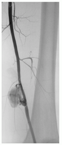
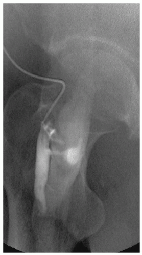
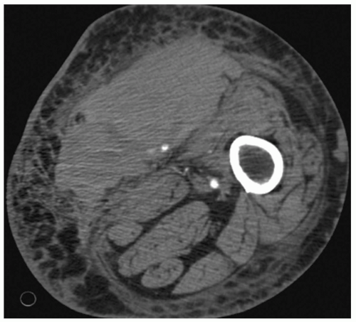
The arterial wall is composed of three anatomic layers-the intima, media, and adventia-and vascular injury can affect one or all vessel layers. Blunt force trauma seen with crush injury can lead to vessel contusion. Hematoma formation occurs between the intima and media or the media and adventia and cause compression and partial or complete vessel occlusion. Disruption between the intima and media can also lead to an intimal flap with subsequent dissection and thrombosis. Whenever the intimal layer is disrupted, the highly thrombogenic internal elastic lamina is exposed. When the inner two layers are disrupted but the adventitia is intact, a pseudoaneurysm forms outside the vessel lumen (see 
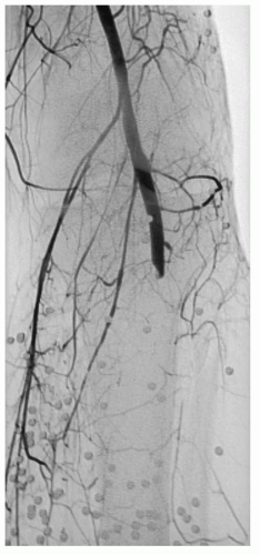
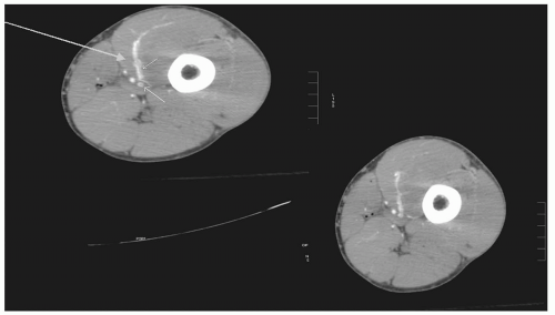
Fig. 1). Stretch injury to the vessel causes disruption between the layers due to shearing forces. These injuries often occur in the setting of posterior knee dislocation with resulting occlusion of the popliteal artery. Vessel lacerations or complete transections are typically the result of penetrating injury from missiles or other projectiles, and less commonly from fractured bone (see Fig. 4). Arteriovenous fistulae (AVFs) commonly result when concomitant arterial and venous injuries occur in close proximity (see Figs. 2 and 6).


Etiology, Injury Mechanism, and Location

The etiology of vascular trauma often depends on both the setting in which injuries are reported and the particular vessel injured. For instance, in the civilian setting at an urban trauma center, penetrating injury accounted for 81% of approximately 360 vascular injuries at the Ben Taub General Hospital in Houston, Texas, from 1981 to 1985.2 Likewise,
recent wartime experience demonstrates that approximately 94% of battlefield vascular injuries (of which 78% were extremity injuries) were the result of penetrating trauma.1 In contrast, injury to the popliteal artery is the result of blunt mechanisms in most reported cases.12,21,22 In their review of the National Trauma Data Bank to evaluate outcomes from popliteal artery injury, Mullenix et al.22 noted that 61% of the injuries were the result of blunt mechanisms, with only 39% occurring from penetrating injury. Likewise, Hossny et al.12 reported that of 39 popliteal artery injuries occurring over a 9-year period, 31 (79%) were the result of blunt trauma.


Figure 1 Superficial femoral artery (SFA) pseudoaneurysm following gunshot wound the thigh.


Figure 2 Common femoral artery (CFA) →femoral vein arteriovenous fistulae (AVF) following penetrating injury to upper thigh.

Penetrating injury most often occurs from gunshot or stab wounds in the civilian setting, and from fragment wounds in combat settings.1,8,11 Surgeons in combat settings also routinely encounter blast injury from improvised explosive devices (IEDs).1 Blunt injury most often results from motor vehicle collisions (MVCs), falls, and crush injury in both civilian and battlefield environments.

Lower extremity vascular injuries are defined as those occurring below the inguinal ligament. The most commonly injured vessel is the femoral artery.6,11 Injuries to the common femoral artery (CFA), profunda femoris, superficial femoral artery (SFA), and popliteal artery both above and below the knee represent the bulk of clinically significant vascular trauma requiring revascularization procedures or surgical exploration (see Table 2). Because there is more redundancy in the blood supply to the calf and foot, with anterior tibial, posterior tibial, and peroneal arteries all supplying this distribution, one or sometimes two of these vessels can be disrupted without loss of distal perfusion and limb ischemia. The one caveat to this point is that significant bleeding from any of these vessels or their branches can lead to elevated compartment pressures in the calf and subsequent compartment syndrome. Trauma and vascular surgeons should maintain a high index of suspicion for the compartment syndrome and intervene with fasciotomy as necessary, as discussed later in this chapter.


TABLE 2 LOCATION OF VESSEL INJURY IN LOWER EXTREMITY, 550 PATIENTS IN 663 COMBINED VASCULAR INJURIES


Vessel


Number of Injuries


Percentage


CFA


55


8.3


Profunda femoral


34


5.1


SFA


238


35.9


Above knee popliteal


129


19.5


Below knee popliteal


86


13.0


Combined above/below knee popliteal


22


3.3


Crural


99


14.9


CFA, common femoral artery; SFA, superficial femoral artery. (Adapted from Hafez HM, Woolgar J, Robbs JV. Lower extremity arterial injury: Results of 550 cases and review of risk factors associated with limb loss. J Vasc Surg. 2001;33(6):1212-1219.)


Vascular injury to the upper extremity is relatively common, representing 30% to 40% of vascular injuries in urban civilian settings.23,24 For the purposes of this discussion, the vessels of the upper extremity are defined as coursing from the axilla to the wrist. Injuries to the subclavian vessels are commonly reported together with those of the axillary vessels, and surgical management and associated injuries are similar.10,25,26 Therefore, axillosubclavian vessel injuries are discussed here as well. The most commonly injured vessel in the upper extremity is the brachial artery with an incidence of approximately 50%, followed by the radial and ulnar arteries (˜25% each), with the least common being the axillary and subclavian arteries (˜3% to 5%).23,24 The most common mechanism for vascular injury in the upper extremity is penetrating trauma.10,23,24,25

Iatrogenic injuries account for an increasing number of peripheral vascular lesions.27 The rise in these injuries has come with the growth of percutaneous therapies for coronary and peripheral arterial disease processes. Although many of these injuries can be managed by noninvasive methods (compression and/or fibrin injection under ultrasonographic guidance, endovascular stent grafts, etc.), some require more extensive management. Although a full discussion of the incidence, etiology, prevention, and management of these injuries is beyond the scope of this chapter, they remain an important diagnostic and treatment challenge for vascular surgeons.


Imaging Techniques


Angiography

Contrast angiography (CA) is considered the gold standard for the evaluation of arterial injury. Proposed for the diagnostic evaluation of extremity vascular trauma as early as the 1960s, arteriography has seen significant advances mostly in the realm of catheter-based therapy (discussed later in this chapter).28,29 As a purely diagnostic modality, angiography has sensitivities of 92% to 96%, specificity above 96%, and 98% reported diagnostic accuracy.30,31,32,33,34 Indications for angiography are mostly based on the clinical findings of the evaluating surgeon and include those mentioned in the preceding text. Some authors use emergency room angiography to quickly triage patients with soft signs of vascular injury.8,35,36 Owing to the potential for multiple lesions within the same vessel or extremity, intraoperative angiography should always be an important part of the surgeon’s armamentarium. This procedure can be performed both before and after repair of identified lesions. In the latter case, after repairing an injured vessel that demonstrated hard signs of injury it may be necessary to perform intraoperative angiography to rule out concomitant distal injury (see Figs. 3 and 4).

Although several scoring systems have been developed to better select patients for CA, such as those with penetrating injury proximity to major vascular structures or relative to injury mechanism, they have not proven to be effective and are not an appropriate substitute for a high index of suspicion following a thorough examination.37,38,39 For instance, it was historically believed that posterior knee dislocations always warranted angiographic investigation because of a high rate of popliteal artery injury. However, because of the low incidence (<16%) of vessel damage in this patient cohort with very few of these injuries of clinical significance, angiogram is only indicated on a selective basis-that is, where PE findings are abnormal.38,40 This is also true for penetrating injury proximity to vascular structures, which is a poor indicator for positive angiography.39


Figure 3 Intraoperative angiography in patient with multiple vascular injuries to a single limb following gunshot wound.


Figure 4 Popliteal artery injury in patient in preceding figure following shotgun blast. Multiple pellets lodged in lower extremity.


Duplex

Duplex ultrasonography can also play a crucial role in evaluating patients with vascular injuries. Although duplex is somewhat limited by operator dependence, several studies have shown that it is a valuable diagnostic tool. In a study of 198 patients with 319 potential injuries, Bynoe et al.41 reported a sensitivity of 95%, a specificity of 99%, and an overall accuracy of 98%. In centers without computed tomography (CT) or angiography readily available, or in-theater forward combat hospitals, ultrasonography may be an invaluable imaging option.


Computed Tomography with Angiogram

Most investigations of the utility of CTA in evaluating vascular injury have focused on either injuries to the neck vessels or those in the lower extremity. For the
purposes of this chapter, the discussion will be centered on the latter. With the advent of improved CT scans, particularly the multidetector contrast-enhanced spiral scanners, detection of vascular injuries has vastly improved. In fact, CTA is becoming a viable alternative to angiography for the detection of arterial injury, particularly in those patients without hard signs. Lesions that can routinely be diagnosed include intimal dissections, pseudoaneurysms, AVFs, thromboses and/or occlusions, hematomas, and laceration with active bleeding as evidenced by contrast extravasation (see Figs. 5 and 6). Occasionally, CTA may be nondiagnostic due to significant artifact from bullet fragments or other foreign bodies.42 Reported sensitivities range from 90% to 100%, with specificities of 98% to 100% (see Table 3). However, some of the recent reports lack comparison to a gold standard (operative exploration or CA) and most are limited by inadequate or short-term follow-up. The sensitivity of CTA in detecting small, although clinically significant lesions such as intimal flaps—particularly those around the knee—is unknown. Large, prospective studies are needed to validate the use of CTA as a substitute for angiography.


Figure 5 Large thigh hematoma with contrast extravastion from branch of superficial femoral artery (SFA).


Figure 6 Contrast extravasation (large arrow) from superficial femoral artery (SFA) injury with arteriovenous fistulae (AVF) (note contrast filling vein, small arrows).


Magnetic Resonance Angiography

Magnetic resonance imaging (MRI) or magnetic resonance angiography (MRA) is used less frequently in the acute setting. Metal implants such as cerebral aneurysm clips and cardiac pacemakers preclude the use of MRI, and it may be difficult to ascertain whether patients have such devices in the acute setting. Many orthopaedic fixation devices are also contraindications for MRI. Additionally, MRI typically takes longer and requires more cooperation from the patient than does CT, which makes it less favorable to use with acutely injured or combative patients. It may also be less effective than CT, as one group of authors showed in a randomized controlled trial that it was not as accurate as CTA in evaluating nontraumatic limb ischemia.47 The ability to discriminate infrapopliteal lesions is limited, and MRA is not as accurate as CA in identifying clinically significant distal lesions in the lower extremity.48 Despite these drawbacks, MRA can provide highly detailed images in the hemodynamically stable patient. In the event that angiography is indicated, but the patient’s renal insufficiency precludes the use of iodinated contrast, MRA may be a good option.


Management


Temporizing Methods

If brisk arterial bleeding is encountered in the primary survey of a trauma patient, digital or manual compression is typically the first method employed for hemorrhage control. In the event of a penetrating injury associated
with brisk bleeding, a Foley catheter can be inserted into the wound tract and inflated to provide compression and partial occlusion. Depending on the setting, tourniquets may be employed for temporary vascular occlusion. These devices are often appropriate in the setting of traumatic amputation or when triaging patients who are in shock at sites a considerable distance from definitive care. It should be noted that tourniquets worsen the ischemic insult, and prophylactic fasciotomy is often routinely used for limbs with prolonged ischemia times. Blindly applying vascular clamps is not advised, as this can cause damage to both the vessel and associated structures.


TABLE 3 THE UTILITY OF COMPUTED TOMOGRAPHY WITH ANGIOGRAM (CTA) IN IDENTIFYING PERIPHERAL ARTERIAL INJURY


Author


Year


Number of CT Scans


Sensitivity (%)


Specificity (%)


Missed Injuries


Mean Follow-up (months)


Sotoetal.43


1999


45


90.0


100.0


NR


NR


Sotoetal.44


2001


139


95.1


98.7


NR


NR


Busquets et al.45


2004


95


100.0


NR


0


8.0


Inaba et al.46


2006


62


100.0


100.0


0


1.5


NR, not reported.


Ligation

Although obviously suboptimal for restoring tissue oxygenation to the affected limb, vessel ligation may be a necessary life-saving option in certain settings. Battlefield or forward theater triage units may perform such maneuvers when definitive vascular surgical repair is not available. This may also be true in the civilian setting where a patient in hemorrhagic shock requires immediate attention to control bleeding before transfer to a Level I trauma center. If vessel ligation is required, it is always advisable to tag the distal end of the vessel and tie it off as well. There are several reasons for this practice. First, significant back-bleeding can occur through collateral vessels and lead to continued shock. Second, when significant blast injury with major tissue disruption occurs, the vessel ends may be more readily identifiable early in the injury evolution. Additionally, muscular arteries have a tendency to contract, requiring extensive dissection during future attempts at repair. It is therefore advisable to make both ends of the ligated vessel readily identifiable for the treating surgeon.

Simple ligation can typically be performed for small distal branches such as tibial arteries in the leg or the radial artery in the forearm without untoward consequences.9,49,50,51 This strategy requires at least one open vessel below the knee or elbow and no evidence of distal ischemia.49 In fact, this operative strategy has been advocated as more cost effective with outcomes equivalent to repair.51


Intravascular Shunt

Temporary intravascular shunts (TIVSs) have been one of the mainstays of trauma surgery for decades.52,53 Providing critical limb perfusion in the case of significant vascular disruption, intravascular shunts are typically used in proximal locations where injury to a major vessel leads to tissue ischemia due to the absence of adequate collateral circulation. This is particularly true at the CFA, SFA and popliteal artery in the lower extremity, and at the subclavian and axillary arteries in the upper extremity (see Fig. 7). Shunts tend to have lower patency rates in smaller, distal vessels.54 Most authors do not advocate prolonged shunting, although case reports have demonstrated shunt patency up to 24 hours.55 Temporary shunts can be invaluable temporizing measures before transfer to a more specialized center for definitive management or before reduction and repair of associated orthopaedic injury.

There are some important principles to keep in mind when employing temporary shunts. First, the tubing must be noncompressible so that it does not collapse when secured proximally and distally within the artery. Commercially available Sundt and Argyle carotid shunts are good options. In the event that these devices are not readily available, sterile, rigid plastic tubing such as intravenous tubing often works well, as do endotracheal suction catheters. Second, the shunt should be sized accordingly, with a diameter slightly smaller than the injured artery. Too large a catheter may cause further intimal damage if it is forcibly passed into the vessel. Likewise, if the catheter is too small, distal flow will be further compromised. Rummel tourniquets with umbilical tape-which is relatively atraumatic to vessel architecture-are a favored technique to secure the shunt within the damaged vessel (Fig. 7). Shunt patency is most dependent on duration of in situ shunting, and systemic heparinization is not required.56,57 Thrombectomy is often necessary, and most authors advocate local intraluminal heparin.52,53,54,55,57,58,59 Outcomes with TIVS are typically good, with significantly fewer complications in shunted versus nonshunted limbs and high limb-salvage rates.12,57,59

Only gold members can continue reading. Log In or Register to continue

Stay updated, free articles. Join our Telegram channel

Oct 17, 2016 | Posted by in MANUAL THERAPIST | Comments Off on Diagnosis and Management of Extremity Vascular Injuries

Full access? Get Clinical Tree

Get Clinical Tree app for offline access