10 Deep dry needling of the hip, pelvis and thigh muscles
Introduction
Pelvic pain is not uncommon in both the male and female population. Pain in the abdominal or pelvic regions that lasts 6 months or more and is not cyclic, is termed chronic pelvic pain (CPP). The American College of Obstetricians and Gynecologists specifies the location of CPP in the pelvis, abdominal wall at or below the umbilicus, lumbar, sacral and buttock regions (Vercellini et al. 2009). Approximately 90% of women with pelvic pain, interstitial cystitis or incontinence have painful trigger points (TrPs) in several locations including the pelvic floor, gluteal and abdominal muscles (Weiss 2001, Wesselmann 2001, Fitzgerald & Kotarinos 2003). The prevalence of CPP is estimated to be 14.7% of women between 18–50 years of age in the USA (Mathias et al. 1996).
Men with chronic prostatitis (CP) and chronic pelvic pain syndrome (CPPS) presented with pain in the scrotal, perineal, inguinal and bladder area in 54% of cases and had tender myofascial palpation in 88% of cases (Zermann et al. 2001). Berger et al. (2007) noted that in men with CPPS, pain was not always limited to the prostate. Anderson et al. (2009) further reported that puborectalis, pubococcygeus and rectus abdominus TrPs reproduced penile pain 75% of the time and external oblique TrPs elicited suprapubic, groin and testicular pain in 80% of the cases.
The European Association of Urology and the Society of Obstetricians and Gynaecologists of Canada recommend that TrPs should be considered in the diagnosis of pelvic pain (Jarrell et al. 2005, Fall et al. 2019). Giamberardino et al. (1999) reported that patients with visceral pain and hyperalgesia are likely to have clinically relevant abdominal TrPs. Myofascial pain was identified in 78.5% of patients with interstitial cystitis and 67.9% of those patients had six or more identifiable TrPs. The most common TrPs were found in the obturator internus, puborectalis, arcus tendineus and iliococcygeus muscles (Bassaly et al. 2010). Specific TrPs were identified in the levator ani muscles in 22% and in the piriformis muscles in 14% of women with CPP.
The International Continence Society (ICS) and the European Association of Urology suggest two categories of pelvic pain: those of muscular origin and pelvic floor muscle spasm (Abrams et al. 2003, Fall et al. 2010); however, other TrPs in the abdominal, gluteal, obturator internus, iliacus, psoas, quadratus lumborum and lumbar multifidi muscles can, and frequently do, refer to the pelvic floor muscles, perineum, vagina, labia, clitoris, scrotum and penis (King-Baker 1993, King & Goddard 1994, Segura et al. 1979, Doggweiler-Wiygul 2004, Doggweiler-Wiygul & Wiygul 2002, Zermann et al. 1999, Prendergast & Weiss 2003).
Deactivation of myofascial TrPs has been supported in the literature to be effective in treating urinary incontinence, chronic prostatitis, chronic pelvic pain, sacroiliac joint dysfunction, dyspareunia, interstitial cystitis, irritable bowel syndrome, levator ani syndrome, and high tone pelvic floor (Lukban et al. 2001, Weiss 2001, Oyama et al. 2004, Anderson et al. 2005, Riot et al. 2004, Tu et al. 2006). A multicenter feasibility study confirmed that TrP therapy is effective in treating CPPS (Fitzgerald 2009). As part of the treatment plan, it is essential to assess the tone of the pelvic floor musculature by a qualified professional when treatment includes dry needling (DN). Chronic pelvic pain can be secondary to low or high tone pelvic musculature. Stability may be compromised if a low tone pelvic floor is deactivated, which may require the additional use of external support until such time the motor control is considered sufficient (Longbottom 2009).
Common pain characteristics are seen with visceral pain originating from pelvic organs and myofascial pain originating from TrPs. The segmental spinal root should also be considered in treatment. Muscle or fascia innervated by the 12th thoracic to the 4th lumbar segments can refer pain to the lower abdomen, iliopsoas, quadratus lumborum, piriformis and obturator internus muscles. The 10th thoracic through the 4th sacral segments supply innervation to the reproductive organs, abdominal wall, low back, thighs and the pelvic floor (Baker 1993, Brookhoff & Bennett 2006, Longbottom 2009).
The aforementioned evidence supports that treatment of TrPs of the pelvic floor and its related structures is an integral part of any therapy regimen. It should be noted that deactivation of TrPs is insufficient if not followed by an appropriate exercise program, which may include specific stretching, strengthening, and motor control training (Edwards & Knowles 2003, Longbottom 2009).
Dry needling of the abdominal, hip, pelvis, and thigh muscles
As in the other chapters, referred pain patterns are mostly based on the descriptions by Simons et al. (1999) with substitutions and additions by Dalmau-Carola (2005) and Longbottom (2009).
Abdominal wall muscles
The rectus abdominus, external and internal obliques muscles are discussed in Chapter 9. The transverse abdominal muscle cannot be needled in isolation of the other abdominal muscles.
Hip muscles
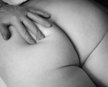
Gluteus maximus muscle
• Anatomy: The muscle originates at the posterior aspect of the ilium, the lower part of the sacrum and coccyx inferior and lateral across the greater trochanter to the iliotibial band of the tensor fascia lata and the gluteal tuberosity.
• Function: Hip extension, lateral rotation and stabilization of the iliotibial tract.
• Innervation: Inferior gluteal nerve from L5, S1 and S2.
• Referred pain: Pain referral is along the inferior or inferior-lateral aspect of the sacrum, the gluteal fold, or insertion along the iliotibial tract. TrPs in the gluteus maximus muscle can imitate sacroiliac pain.
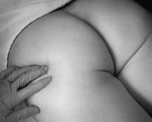
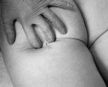
• Needling technique: The patient is prone with a pillow under the abdomen or side lying. The muscle is needled with flat palpation perpendicular to the muscle along the area of the TrP (Figure 10.1). Strong depression of the subcutaneous tissue is required to reduce the distance from the skin to the muscle.
• Precautions: Avoid needling the sciatic nerve. Depth of penetration is dependent on the amount of adipose tissue.
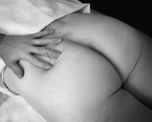
Gluteus medius muscle
• Anatomy: The muscle is found between the gluteus maximus and tensor fascia latae. It originates between the posterior and anterior gluteal lines of the ilium and inserts on the lateral border of the greater trochanter. A bursa lies under the tendinous portion over the surface of the trochanter.
• Function: Hip abduction and medial rotation. Insufficiency of this muscle results in a positive Trendelenburg test.
• Innervation: Superior gluteal nerve from L4, L5 and S1.
• Referred pain: TrPs may be found throughout the entire muscle with referral to the sacroiliac joint, gluteal and lumbosacral regions, and along the iliotibial tract, gluteal region, posterior thigh and posterior lower leg. It is not possible to separate referred pain patterns from the gluteus minimus muscle in the area where the two muscles overlap.
• Needling technique: The patient is prone or side lying. The muscle is needled with flat palpation perpendicular to the muscle along the contour of the iliac crest. Strong depression of the subcutaneous tissue is required to reduce the distance from the skin to the muscle. Needle contact at the periosteum is common (Figure 10.2).
• Precautions: Avoid needling the sciatic nerve. There are also deep branches of the superior gluteal vessels and nerve between the medius and minimus which should be not needled. Depth of penetration is dependent on the amount of adipose tissue.
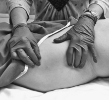
Gluteus minimus muscle
• Anatomy: The muscle is found deep to the gluteus medius. It originates between the anterior and inferior gluteal lines of the anterior aspect of the ilium and inserts on the anterior aspect of the greater trochanter. It also has a bursa between the tendon and the insertion at the greater trochanter.
• Function: Hip abduction and medial rotation. Insufficiency of this muscle along with the gluteus medius results in a positive Trendelenburg test. Supports the body in single leg stance with the tensor fascia latae.
• Innervation: Superior gluteal nerve from L4, L5 and S1.
• Referred pain: Referred pain from the gluteus minimus muscle is into the iliotibial tract, gluteal region, posterior thigh and posterior one third of the lower leg. It is not possible to separate referred pain patterns from the gluteus medius muscle in the area where the two muscles overlap.
• Needling technique: The patient is prone or side lying. The muscle is needled with flat palpation perpendicular to the muscle along the contour of the iliac crest. Strong depression of the subcutaneous tissue is required to reduce the distance from the skin to the muscle. Needle contact at the periosteum is common (Figure 10.3).
• Precautions: There are deep branches of the superior gluteal vessels and nerve between the medius and minimus which should be not needled. Depth of penetration is dependent on the amount of adipose tissue.
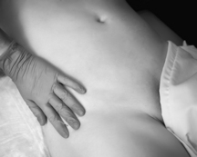
Tensor fascia latae muscle
• Anatomy: The muscle originates from the anterior outer aspect of the iliac crest and the anterior superior iliac spine (ASIS), between the gluteus medius and sartorius; and from the deep surface of the fascia lata. It inserts between the two layers of the iliotibial band of the fascia latae of the middle and upper thirds of the thigh.
• Function: Via the iliotibial tract it extends the knee with lateral rotation of the leg, assists in flexion, abduction and medial rotation of the hip. Pelvic stabilization and posture control are its main functions.
• Innervation: Superior gluteal nerve from L4, L5 and S1.
• Referred pain: The referred pain from this muscle travels along the iliotibial tract, gluteal region or posterior lateral thigh.
• Needling technique: The patient is supine or side lying. The muscle is needled with flat palpation perpendicular to the muscle (Figure 10.4).
Obturator internus muscle
• Anatomy: The obturator internus muscle has a broad attachment to the anterior lateral wall of the inner pelvic brim, the rim of the obturator foramen and the obturator membrane, covering most of the obturator foramen. The pelvic surface of the obturator internus and its fascia form the anterior lateral wall of the true pelvis. The muscle exits the pelvis through the lesser sciatic foramen then makes a right angle turn, posterior to the hip joint capsule, attaching to the anterior medial surface of the greater trochanter in close proximity to the gemelli muscles.
• Function: The primary function of the obturator internus is to laterally rotate the hip joint when the thigh is extended and assist with hip abduction when the hip is flexed to 90°.
• Innervation: The muscle is supplied by the nerve to the obturator internus from L5 and S1
• Referred pain: The obturator internus muscle refers pain to the vagina, anococcygeal region and the posterior thigh. The patient may also have a perception of fullness in the rectum.
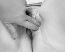
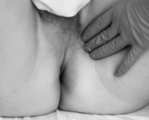
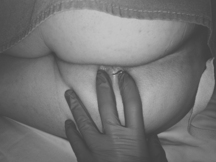
• Needling technique: In the lithotomy position, palpate the inferior border of the pubic ramus and the insertion of the adductor longus tendon. Just lateral palpate toward the obturator foramen. Angle the needle perpendicular and lateral to the muscle surface directly into the TrP taut band identified with internal or external palpation (Figure 10.5 – female and Figure 10.6 – male). Another option is side-lying on the involved side with hip and knee flexion. Palpate the ischial tuberosity. Place the fingers medially around the bony prominence then angle the needle perpendicular to the muscle surface with a slight anterior superior angle directly into the TrP taut band identified with internal or external palpation (Figure 10.7).
• Precautions: Avoid needling into the Pudendal (Alcock’s) canal containing the pudendal and obturator nerve and vessels.
Obturator externus/gemellus inferior and superior muscles
• Anatomy: The obturator externus is a flat triangular muscle that covers the external surface of the obturator membrane and adjacent bone of the ischial and pubic rami laterally and upward to the trochanteric fossa of the femur. The superior gemellus originates at the spine of the ischium and inferior from the ischial tuberosity. Collectively they insert into the medial surface of the greater trochanter of the femur. Functionally, the gemelli muscles can be considered to be part of the obturator internus muscle and these muscles may actually be fused together (Honma 1998).
• Function: Lateral rotation of the hip as well as stability of the hip and pelvis.
• Innervation: The obturator externus is innervated by the posterior branch of the obturator nerve from L3-L4, the gemellus superior by branches of the obturator internus nerve form L5–S2, and the gemellus inferior by branches of the quadratus femoris nerve from L4–S1.
• Referred pain: Referred pain patterns from this muscle are not described in detail, but are generally thought to include the proximal posterior thigh, hip and groin as well as sciatic type referral.
• Needling technique: The patient is positioned comfortably prone. Palpate the greater trochanter medially and insert the needle perpendicular to muscle surface directly into the TrP taut band identified by palpation. The obturator externus is found deeper and more posterior to the trochanter (Figure 10.8).
Quadratus femoris muscle
• Anatomy: This is a flat quadrilateral muscle that originates at the upper part of the external aspect of the ischial tuberosity and inserts above the middle of the trochanteric crest of the femur.
• Function: Lateral rotation of the thigh
• Innervation: Branches from the quadratus femoris nerve from L5 and S1.
• Referred pain: Referred pain patterns from this muscle are not described in detail, but are generally thought to include the proximal posterior thigh, hip and groin as well as sciatic type referral.
• Needling technique: The patient is positioned comfortably prone. Palpate the greater trochanter and the ischial tuberosity. The needle can be inserted perpendicular to the muscle surface at the trochanter or just medial to the ischial tuberosity running parallel to the sciatic nerve, directly into the TrP taut band identified by palpation (Figure 10.9).
Stay updated, free articles. Join our Telegram channel

Full access? Get Clinical Tree


