Decompression of the Ulnar Nerve at Guyon Canal
Harris Gellman
Patrick Owens
DEFINITION
The site of compression must be identified to determine the appropriate treatment for symptoms of ulnar nerve dysfunction. Guyon canal at the wrist is the second most common site of ulnar nerve entrapment.
Symptoms may be purely motor, purely sensory, or mixed, depending on the site and cause of compression.
ANATOMY
In the distal half of the forearm, the ulnar nerve is joined on its lateral side by the ulnar artery. Proximal to the wrist, the nerve gives off a large dorsal sensory branch, which supplies sensation to the dorsum of the wrist and the ulnar side of the hand. The ulnar nerve continues into the hand through Guyon canal.
Guyon canal is a triangular canal at the base of the ulnar side of the palm. It is 4 cm in length, extending from the proximal edge of the palmar carpal ligament to the fibrous edge of the hypothenar muscles.4 The space functions as a physiologic tunnel with discrete anatomic landmarks (FIG 1A).
Both the ulnar nerve and artery pass through the canal to enter the hand.
The dorsal cutaneous branch of the ulnar nerve usually branches before the nerve enters Guyon canal.
It is bordered laterally by the hook of the hamate and the transverse carpal ligament. The medial wall is formed by the pisiform and the attachments of the pisohamate ligament.
Dividing the tunnel into three zones helps in correlating the clinical symptoms with the specific pathologic cause4,13 (FIG 1B).
Zone 1, about 3 cm in length, is the area proximal to the bifurcation of the ulnar nerve into motor and sensory branches. Compression in zone 1 results in combined motor and sensory loss. It is most commonly caused by a fracture of the hook of the hamate or a ganglion cyst.
Zones 2 and 3 are located next to each other, from the point where the ulnar nerve divides into a superficial or sensory branch and a deep motor branch to the region just beyond the fibrous arch of the hypothenar muscles.
Zone 2 encompasses the motor branch of the nerve, located in the dorsoradial portion of the tunnel. The deep motor branch, along with the deep branch of the ulnar artery, passes between the abductor digiti quinti and the flexor digiti quinti brevis, perforating the opponens digiti quinti. The motor branch then follows the deep volar arch across the palm to innervate the interossei.
The nerve supplies the three intrinsic muscles of the small finger, the third and fourth lumbricals, the volar and dorsal interossei, the adductor pollicis, and the deep head of the flexor pollicis brevis.
Compression in this area causes pure motor loss to all of the ulnar-innervated muscles in the hand. Ganglions from the pisotriquetral joint and fractures of the hook of the hamate are the most common etiologic factors (FIG 1C). Due to the nerve’s proximity to the hamate, it is unfortunately easy to damage the nerve while excising the hook of the hamate.
Zone 3, located ulnar to zone 2, encompasses the superficial or sensory branch of the bifurcated ulnar nerve. Compression here causes sensory loss to the hypothenar eminence, the small finger, and part of the ring finger but does not usually cause motor deficits. Common causes are aneurysm of the ulnar artery, thrombosis, and synovial inflammation.
The superficial branch of the ulnar nerve in Guyon canal supplies the palmaris brevis and the skin of the hypothenar eminence and forms the digital nerves to the small and ulnar side of the ring finger.
Two specific nerve anomalies can confuse the diagnosis.
Martin-Gruber anastomosis in the forearm: Fibers that supply the intrinsic muscles are carried in the median nerve to the middle of the forearm, where they leave the median nerve to join the ulnar nerve. Functioning intrinsic muscles can be observed when the ulnar nerve is injured proximal to this anastomosis.
Riche-Cannieu anastomosis: The median and ulnar nerves are connected in the palm. Even with an injury at the wrist, some intrinsic function remains.
PATHOGENESIS
Causative factors of compression or injury of the ulnar nerve in Guyon canal include repeated blunt trauma from power tools and gripping or hammering with the palm of the hand, which may result in thrombosis or aneurysm of the ulnar artery compressing the nerve (hypothenar hammer syndrome)2,6,10 (FIG 2A,B; Table 1). Direct pressure on the ulnar nerve may occur during activities such as cycling.
Fractures of the hook of the hamate can impinge on the nerve.
Idiopathic compression may occur secondary to thickening of the proximal fibrous ligament at the entrance to the canal.
Compression also may occur as a result of swelling after distal radius fracture.
Compression of the ulnar nerve at Guyon canal also has been shown to occur in conjunction with carpal tunnel syndrome. It typically resolves after surgical decompression of the carpal canal.9,16
Other etiologies include tumors such as ganglia or lipomas (FIG 2C,D), anomalous muscle bellies,8,15 or hypertrophy of the palmaris brevis.
Ganglia and other soft tissue masses are responsible for 32% to 48% of cases of ulnar tunnel syndrome. Another 16% of cases are due to muscle anomalies.12
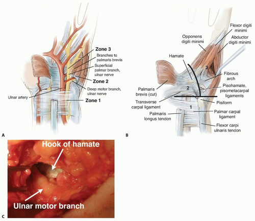
FIG 1 • A. Anatomic landmarks of the distal ulnar tunnel (Guyon canal). Zone 1: ulnar nerve in the region proximal to the bifurcation. Zone 2: ulnar nerve motor segment following bifurcation. Zone 3: ulnar nerve sensory segment distal to the bifurcation. B. Location of the three zones. Zone 1 is proximal to the bifurcation; zone 2 encompasses the motor branch; zone 3 is the region surrounding the sensory branch. C. The proximity of the motor branch of the ulnar nerve to the hook of the hamate as seen during excision of the hook.
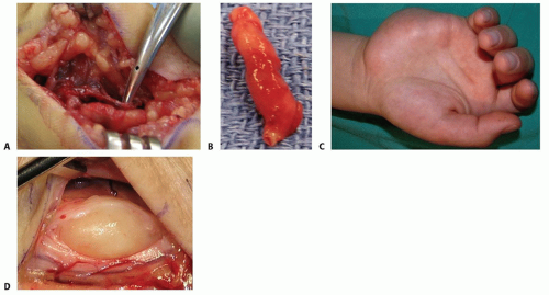
FIG 2 • A. Ulnar artery thrombosis. B. Resected thrombosed segment. C. Hypothenar mass as a cause of compression of the ulnar nerve at the wrist. D. Lipoma causing compression of the nerve.


