Clinodactyly and Camptodactyly
CASE PRESENTATION
A 6-month-old infant presents with bilateral flexion contractures of the small finger proximal interphalangeal (PIP) joints: 30 degrees on the left and 60 degrees on the right by passive exam. Active motion is more limited than passive motion. The parents think this has been present since birth but thought it was a normal, clenched fisting posture of newborns. There is no family history of congenital hand differences. No associated conditions have been noted by the primary care physician on exam.
CLINICAL QUESTIONS
What are clinodactyly and camptodactyly?
What is a delta phalanx? Bracket epiphysis?
What is the normal anatomy around the PIP joint of a finger?
What is the abnormal anatomy in clinodactyly and camptodactyly?
What are the associated conditions and syndromes?
When is nonoperative treatment indicated?
What are the indications for surgery?
Is surgery commonly performed?
What techniques are used to correct camptodactyly of the fingers and clinodactyly of the fingers and thumbs?
What are the downsides and complications of surgery?
THE FUNDAMENTALS
Do not let what you cannot do interfere with what you can do.
—John Wooden
“Crooked” fingers and thumbs are normal to a certain degree as long as they do not interfere with adjacent digital and/or overall hand function. Every patient will have straighter fingers when they actively flex into the palm than when passive alignment is checked by tenodesis. Check yourself now and see what you find out about your passive and active alignment differences. Most if not all nonpathologic, digital malalignment and malrotation in people are symmetric. So, always examine the other side to determine if what you and they are seeing is really abnormal. In general, <30 degrees of a flexion contracture at the PIP joint or 15 degrees of radioulnar deviation does not compromise hand function. The patient’s and parents’ aesthetic perception is another issue altogether.
Grasp and power grip on the ulnar side are important hand functions that are not impaired by a flexion contracture of the interphalangeal (IP) joint. Digital release is well maintained by compensatory hyperextension of the metacarpophalangeal (MCP) and distal interphalangeal (DIP) joints, especially in ligamentously lax young patients.
Etiology and Epidemiology
Clinodactyly
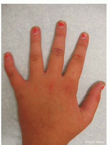
Clinodactyly by definition is >10 degrees of digital deviation in the radioulnar plane. This is usually due to abnormal bony anatomy. It can involve any digit but is most common in the small finger due to an abnormally shaped middle phalanx. Asymmetric longitudinal growth of the physis results in digital malalignment, usually radial deviation of the small finger. Up to 1% of the population will have clinodactyly of the small finger (Figure 6-1). Bilateral involvement is common. There is an autosomal dominant inheritance pattern in isolated, familial clinodactyly of the small finger.1
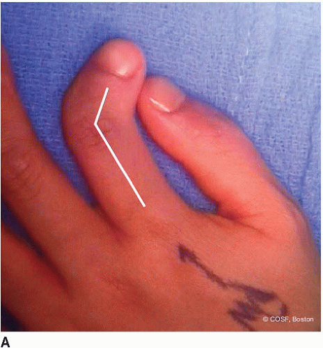
Clinodactyly of the index finger is a unique entity that presents at birth with marked deformity (Figure 6-2). It is often unilateral, with male predominance. It is commonly associated with brachydactyly and a hypoplastic trapezoidal or triangular middle phalanx. It is not associated with systemic anomalies or mental retardation. The index finger is usually radially deviated.2
Triphalangeal thumb is a unique form of clinodactyly in which the “extra bone” is triangular or trapezoidal shaped. This delta phalanx leads to malalignment of the thumb and, if severe, can impair pinch. Too much length with a rectangular extra phalanx can also impair thumb
function. Inheritance is autosomal dominant. It is seen with other thumb malformations, such as thumb polydactyly. It is usually not seen with other systemic conditions or syndromes.
function. Inheritance is autosomal dominant. It is seen with other thumb malformations, such as thumb polydactyly. It is usually not seen with other systemic conditions or syndromes.
 FIGURE 6-1 Mild, nonprogressive clinodactyly with radial deviation of small finger due to trapezoidal-shaped middle phalanx. |
Associated syndromes and conditions are common. Delta phalanges and bracket epiphyses are often a part of other, more complex hand conditions (see Chapter 5). They are seen as a frequent part of chromosomal, central nervous system, and craniofacial abnormalities (Table 6.1). Many conditions that include symptoms of impaired intelligence have clinodactyly as part of their clinical spectrum. In utero ultrasound screening for Down syndrome includes clinodactyly as one of the diagnostic criteria. Clinodactyly of the thumb is seen most often with Rubinstein-Taybi syndrome, Apert syndrome, and dystrophic dysplasia (hitchhiker thumb).
 FIGURE 6-2 A: AP clinical view of index finger clinodactyly. Arrows outline deformity due to middle phalangeal delta phalanx. |
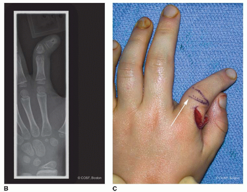
Clinodactyly can be congenital (as above) or acquired. Acquired situations are due to physeal changes from trauma, infection, and benign tumors (intra-articular osteochondroma enchondroma) (Figure 6-3).3
Camptodactyly
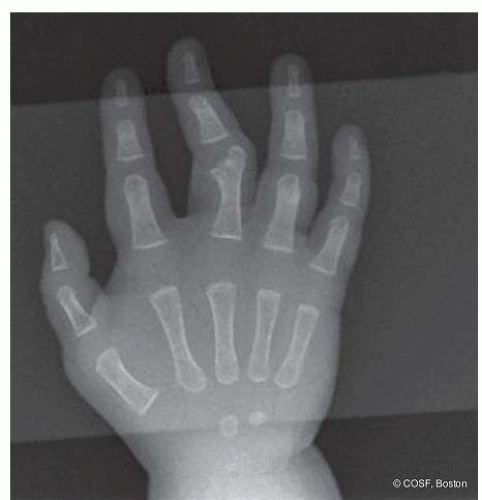
Camptodactyly by definition is a flexion posture or contracture of the PIP joint. Like clinodactyly, it usually involves the small finger and is often bilateral, though typically asymmetric (Figure 6-4). There is an autosomal dominant inheritance pattern of variable penetrance and expressivity with isolated small finger camptodactyly. Less than 1% of people have camptodactyly. Unlike clinodactyly, camptodactyly is usually secondary to soft tissue abnormalities. The most commonly implicated anatomic structures are the flexor digitorum superficialis (FDS) and the lumbrical. The abnormal volar static and dynamic forces lead to skin, fascial, volar plate, check rein ligament, and collateral ligament tightness. Eventually bone and joint deformity develops (Figure 6-5). Camptodactyly often presents during periods of accelerated growth. Since infants usually double their size in the first 6 months and triple their size in
the first year, this is a common time for presentation. Similarly, you can almost hear teenagers grow as they eat their way through their families’ refrigerators, so adolescents are the second-most common presenters of this condition to the office.
the first year, this is a common time for presentation. Similarly, you can almost hear teenagers grow as they eat their way through their families’ refrigerators, so adolescents are the second-most common presenters of this condition to the office.
Camptodactyly is seen with other orthopaedic or generalized conditions. The most common orthopaedic condition with camptodactyly is arthrogryposis, usually involving multiple digits. Various types of skeletal dysplasias can have camptodactyly. Beal arachnodactyly is another syndrome that has associated PIP joint flexion contractures. Marfan syndrome has PIP joint flexion posturing. The extreme form of a flexion contracture at the PIP joint is symphalangism. Like clinodactyly, the associated syndromic conditions include genetic and craniofacial abnormalities (Table 6.2). There is a unique genetic condition described in Saudi Arabian families, the camptodactylyarthropathy-coxa vara-pericarditis (CACP) syndrome. It is an autosomal recessive disorder caused by mutations in the proteoglycan 4 (PRG4) gene. Manifestations vary across families as well as between affected individuals from the same family, with digital camptodactyly and arthropathy of the knees the most ubiquitous, while pericarditis is evident in only one-fifth of all reported cases.4,5
Table 6.1 Representative syndromes associated with clinodactyly (not exhaustive but representative) | |||||
|---|---|---|---|---|---|
|
Clinical Evaluation
About the only thing that comes to us without effort is old age.
—Gloria Pitzer
Clinodactyly
For both clinodactyly and camptodactyly, careful physical examination and thorough patient/parental counseling are critically important. In clinodactyly, measurement of the angular deformity from proximal to distal aspects of the digit through the point of maximum deformity is recorded. This is remeasured with each visit to assess progression, or lack thereof, with growth. The usual pathology is asymmetric longitudinal growth of the middle phalanx physis in the finger and proximal phalanx in the thumb. The quality of the volar digital flexion creases at the IP joints provides insight into the underlying bony architecture. In clinodactyly, the PIP and DIP converge on the radial side rather than remain parallel (Figure 6-6


Stay updated, free articles. Join our Telegram channel

Full access? Get Clinical Tree


