Fig. 2.1
L2, L3 and L4 spinal roots’ contribution to femoral nerves
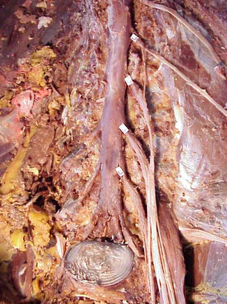
Fig. 2.2
Component of femoral nerves—L2, L3 and L4 spinal roots
2.2.1 Anatomical Study of Knee Jerk Reflex Related Nerve: Femoral Nerve
Firstly, femoral nerve was tracked to its’ central terminal, and the contribution of L2, L3 and L4 spinal nerve roots to the femoral nerve was observed. The diameter of each root at the point of intervertebral foramen was measured, and the diameter of each branch contributing to femoral nerve was recorded as well. Then, a picture was taken after the exposure of conus medullaris and cauda equina, as the end of spinal cord cone was marked with a needle, the anterior-posterior and sequence relationships of L2–4 and S2~4 anterior and posterior roots were recorded.
2.2.2 Anatomical Study of Achilles Tendon Reflex Related Nerve: Sciatic Nerve
Sciatic nerve is the main nerve extended from sacral plexus. So firstly, L4, L5 and S1 spinal roots’ contribution to sacral plexus was observed and the diameter of each root at the point of intervertebral foramen was measured, and the diameter of each branch contributing to sacral plexus was recorded as well. Then, a picture was taken after the exposure of conus medullaris and cauda equina, as the end of spinal cord cone was marked with a needle, the anterior-posterior position and sequence relationships of L4 ~ S1 and S2~4 anterior and posterior roots were recorded.
2.2.3 Anatomical Study of T11–12 Rib Nerves and Spinal Roots Related to Lower Abdominal Wall Reflex
As spinal dura was opened and medullary conus and T11, T12 and S2–4 spinal roots were exposured, anatomical study about the anterior-posterior position and sequence relationships of T11, T12 and S2–4 spianl roots were performed.
Stastistical analysis: SPSS 10.0 was used for analysis of ventral spinal roots’ horizontal area.
2.3 Results
2.3.1 Anatomical Study of L2–4 Spinal Roots and Femoral Nerve (Related to Knee Jerk Reflex)
L2, L3 and L4 spinal roots are the major part contributing to femoral nerve, and there is a hairliking branch of L1 spinal root to join lumbar plexus and femoral nerve which is too small to be measured. L3 and L4 spinal roots’ contributions (42 % and 39 %) to femoral nerves are great than L2 spinal roots’ (19 %), and there is no statistical difference between L3 and L4 spinal roots’ (Fig. 2.1).
As femoral is not the only nerve originated from L2, L3 and L4 spinal roots, which also form obturator nerve and L4 spinal root will send a branch to join the sacral plexus. Figure 2.2 show how much part of L2, L3 and L4 spinal root contributes to femoral nerve: the proportion of L2 spinal root (86 %) and that of L3 spinal root (82 %) is more than that od L4 spinal root (64), p < 0.05, and there is no statistical difference between that of L3 and L4 spinal roots.
Intradural relative position between L2-4 and S2-4spinal roots: It is observed in 39 sides in 20 specimens that the level of S2~4 anterior spinal roots emenate from spinal cord is higher than the level of L2~4 anterior spinal roots cross meninx, that means and the horizontal area of L3 or L4 anterior spinal roots is more than that of S2~4 anterior spinal roots. Those two levels locate at the same plane is observed in 1 side of 40 sides in 20 specimens.
2.3.2 Anatomical Study of L4–S1 Spinal Roots, Sacral Plexus and Sciatic Nerve (Related to Achilles Tendon Reflex)
Sacral plexus are mainly extended from L4, L5, S1 and S2 spinal roots where L4, L5 and S1 spinal roots’ contribution is greater than S2 spinal root’s. Among L4, L5 and S1 spinal roots, L4 spinal root’s contribution (15 %) is less than L5(46 %) and S1(39 %) spinal roots’ (p < 0.01), and there is no statistical difference between L5 and S1 spinal roots’ contribution (p > 0.05, Figs. 2.3 and 2.4).
Fig. 2.3
L2, L3 and L4 spinal roots’ distribution
Fig. 2.4
L4, L5 and S1 spinal roots’ contribution to sacral plexus
Overlapping length between L4 ~ S1 and S2~4 ventral roots: S2~4 the level of ventral roots emenate from spinal cord located at L1 vertebral body level in 28 sides of 20 specimens and it located at L2 vertebral level in other 12 sides of 20 specimens. The level of S2~4 ventral spinal roots emanate from spinal cord is higher than the level of L4 ~ S1 spinal roots cross dura, that means, the end part of L4 ~ S1 ventral spinal roots and the beginning part of S2–4 is overlapping. The overlapping length between L4 ~ S1 and S2~4 ventral roots is showed in Table 2.1.
Table 2.1
Overlapping length between L4 ~ S1 and S2~4 ventral roots  ±s (Min ~ max) cm
±s (Min ~ max) cmL4 | L5 | S1 | |
|---|---|---|---|
S2 | 10.3 ± 2.3 | 13.0 ± 2.3 | 15.0 ± 3.1 |
(7.1 ~ 13.4) | (10.0 ~ 16.7) | (12.0 ~ 8.9) | |
S3 | 9.7 ± 2.3 | 12.3 ± 2.2 | 14.4 ± 8.2 |
(6.6 ~ 12.3) | (9.5 ~ 15.6) | (11.5 ~ 17.8) | |
S4 | 8.3 ± 2.4 | 10.9 ± 2.3 | 13.0 ± 2.2 |
(4.6 ~ 11.1) | (7.5 ~ 13.6) | (9.5 ~ 15.8) |
2.3.3 Anatomical Study of T11–12 Rib Nerves and Spinal Roots Related to Lower Abdominal Wall Reflex
T11 and T12 spinal roots directly extend into T11 and T12 rib nerves which relate to lower abdominal wall reflex. There is no statistical difference between the horizontal areas of T11(0.70 ± 0.14 mm2), T12(0.61 ± 0.18 mm2) and S3(0.61 ± 0.07 mm2), S4(0.45 ± 0.09 mm2) anterior roots(p > 0.05), but the horizontal areas of T11, T12 anterior roots is significantly less than that of S2 anterior roots(1.12 ± 0.24 mm2), p < 0.05 (Table 2.2).
Table 2.2
 Selective Sacral Rhizotomy: Introducing a Simple Intraoperative Manometric Method
Reconstruction of Bladder Innervation Above the Level of Spinal Cord Injury for Inducing Urination by Abdomen-to-Bladder Reflex Contractions
Reconstruction of Bladder Innervation Below the Level of Spinal Cord Injury for Inducing Urination by Achilles Tendon-to-Bladder Reflex Contractions
Pathological Changes in the Detrusor After Spinal Cord Injury
Reconstruction of Afferent and Efferent Nerve Pathways of the Atonic Bladder
Electrical Stimulated Micturition: Sacral Anterior Root Stimulator + Sacral Deafferentation
Selective Sacral Rhizotomy: Introducing a Simple Intraoperative Manometric Method
Reconstruction of Bladder Innervation Above the Level of Spinal Cord Injury for Inducing Urination by Abdomen-to-Bladder Reflex Contractions
Reconstruction of Bladder Innervation Below the Level of Spinal Cord Injury for Inducing Urination by Achilles Tendon-to-Bladder Reflex Contractions
Pathological Changes in the Detrusor After Spinal Cord Injury
Reconstruction of Afferent and Efferent Nerve Pathways of the Atonic Bladder
Electrical Stimulated Micturition: Sacral Anterior Root Stimulator + Sacral Deafferentation


Horizontal area of L4 ~ S4 ventral spinal roots
Related posts:
Selective Sacral Rhizotomy: Introducing a Simple Intraoperative Manometric Method
Reconstruction of Bladder Innervation Above the Level of Spinal Cord Injury for Inducing Urination by Abdomen-to-Bladder Reflex Contractions
Reconstruction of Bladder Innervation Below the Level of Spinal Cord Injury for Inducing Urination by Achilles Tendon-to-Bladder Reflex Contractions
Pathological Changes in the Detrusor After Spinal Cord Injury
Reconstruction of Afferent and Efferent Nerve Pathways of the Atonic Bladder
Electrical Stimulated Micturition: Sacral Anterior Root Stimulator + Sacral Deafferentation
Stay updated, free articles. Join our Telegram channel
Full access? Get Clinical Tree
