Cardiac Malposition and Heterotaxy
Howard P. Gutgesell
CARDIAC MALPOSITION
The term cardiac malposition implies location of the heart anywhere other than in its usual position in the left hemithorax, or it may describe location of the heart in the left hemithorax when other organs are in abnormal positions, as in situs inversus viscerum. Dextrocardia, levocardia, and mesocardia are general terms that indicate the cardiac position only and do not describe intracardiac anatomy. Dextrocardia denotes a right-sided heart, levocardia a left-sided heart, and mesocardia a midline heart. Situs solitus is the normal or usual arrangement of organs (i.e., heart on left, liver on right, stomach on left). Situs inversus is the mirror image of normal (i.e., heart on right, liver on left, stomach on right). The term heterotaxy designates abnormal arrangements of body organs that differ from the orderly arrangements of situs solitus or situs inversus. Typically, duplication or absence of normally unilateral structures occurs (especially the spleen). The terms situs ambiguus and indeterminate situs are synonymous with heterotaxy. Isomerism indicates presence of paired, mirror-image sets of normally unilateral structures, such as the lungs and atria. Left isomerism refers to the presence of two anatomic left lungs and two left atria, whereas right isomerism implies bilateral right lungs and atria.
Dextrocardia
The incidence of situs inversus is 1 in 8,000 to 1 in 7,000 living persons. Dextrocardia with situs solitus (isolated dextrocardia) occurs less commonly. Incidence estimates are as low as 1 in 29,000.
Dextroversion, dextrorotation, and pivotal dextrocardia describe dextrocardia with situs solitus. Often, the heart appears as if the apex has been swung from the left side of the chest to the right side. The term isolated dextrocardia similarly connotes that the other organs are in their normal locations and that dextrocardia is an isolated finding. Generally, mirror-image dextrocardia is applied to more or less normal hearts in subjects with situs inversus. Dextrocardia resulting from displacement of the heart into the right hemithorax by external causes (pneumothorax, diaphragmatic hernia, or hypoplasia of the right lung) is termed secondary dextrocardia or dextroposition.
Although dextrocardia can be diagnosed by physical examination, usually it is detected by chest roentgenography. The clinical presentation may be that of a newborn with cyanosis, respiratory distress, or heart murmur. In cases of secondary dextrocardia, a chest roentgenogram may be the only diagnostic test necessary (e.g., in pneumothorax). In the absence of such an obvious cause, the initial step in evaluating dextrocardia is to determine the situs of the other viscera. Frequently, a chest roentgenogram is useful in showing the location of the liver and stomach. The situs of the lungs may be inferred from chest films. On the electrocardiogram (ECG), a P vector directed leftward and inferiorly suggests situs solitus of the atria, whereas a rightward P axis suggests situs inversus. The details of visceral situs and intracardiac anatomy can be determined by echocardiography, supplemented by magnetic resonance imaging or angiocardiography.
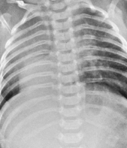
Frequently, dextrocardia in the presence of situs solitus is associated with major intracardiac abnormalities (Fig. 270.1). The most common findings are summarized in Table 270.1. Often, atrioventricular discordance (L-loop ventricles), single ventricle, transposition, and pulmonary stenosis or atresia are present.
The scimitar syndrome is an uncommon but well-described constellation of cardiopulmonary anomalies consisting of dextrocardia, situs solitus of the atria and viscera, hypoplasia of the right lung, anomalous systemic arterial blood supply to the right lung, and anomalous pulmonary venous connection of the right lung to the inferior vena cava. Often, the anomalous pulmonary vein is visible on the chest roentgenogram as a curvilinear shadow in the right lung and resembles a Turkish sword or scimitar (Fig. 270.2).
The incidence of congenital heart disease in subjects with dextrocardia and situs inversus is much lower than that in
subjects with dextrocardia and situs solitus. Although precise determination is not available, the incidence of congenital heart disease may not differ from that in the general population (approximately 8 in 1,000). Cardiac abnormalities found in dextrocardia with situs inversus are summarized in Table 270.1. Atrioventricular discordance and transposition complexes are common findings but occur less frequently than in dextrocardia with situs solitus. Double-outlet right ventricle, pulmonary stenosis or atresia, and ventricular septal defect are present in one-third to two-thirds of reported cases. Usually, the aortic arch is right-sided.
subjects with dextrocardia and situs solitus. Although precise determination is not available, the incidence of congenital heart disease may not differ from that in the general population (approximately 8 in 1,000). Cardiac abnormalities found in dextrocardia with situs inversus are summarized in Table 270.1. Atrioventricular discordance and transposition complexes are common findings but occur less frequently than in dextrocardia with situs solitus. Double-outlet right ventricle, pulmonary stenosis or atresia, and ventricular septal defect are present in one-third to two-thirds of reported cases. Usually, the aortic arch is right-sided.
TABLE 270.1. INCIDENCE OF INTRACARDIAC ABNORMALITIES IN PATIENTS WITH DEXTROCARDIA | |||||||||||||||||
|---|---|---|---|---|---|---|---|---|---|---|---|---|---|---|---|---|---|
|
 FIGURE 270.1. Chest roentgenogram in a neonate with dextrocardia. Echocardiography revealed situs solitus and normal intracardiac anatomy. The heart has shifted to the right, a process probably related to hypoplasia of the right lung.
Stay updated, free articles. Join our Telegram channel
Full access? Get Clinical Tree
 Get Clinical Tree app for offline access

|


