Fig. 61.1
Bizarre parosteal osteocartilaginous proliferation (BPOP). Radiographic and macroscopic features. (a) Bone and cartilaginous lesion of the metacarpal bone. (b) Well-circumscribed osteocartilaginous nodule
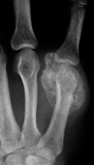
Fig. 61.2
Bizarre parosteal osteocartilaginous proliferation. Plain film, anteroposterior view of the fifth metacarpal bone shows a uniform mineralized well circumscribed lesion in the bone surface
Fig. 61.3
(a) Bizarre parosteal osteocartilaginous proliferation of the middle phalanx (arrow) clearly shows the parosteal location in this radiograph. (b) MRI of same lesion
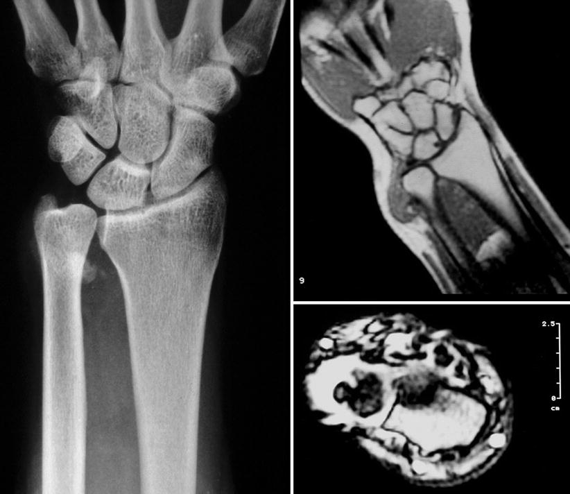
Fig. 61.4
BPOP. X-ray and MRI. Distal ulna well-circumscribed radiodensity
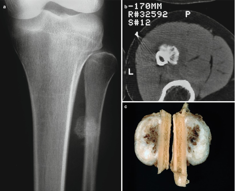
Fig. 61.5


(a) X-ray and (b) CT scan. BPOP attached to the cortex of the fibula. (c) Gross appearance
Related posts:


Stay updated, free articles. Join our Telegram channel
Full access? Get Clinical Tree
