Biomechanics


Biomechanics


image


JOINT ROM


Hip


Rotation


Int


Ext


Adults


35°-40°


35°-40°


Infants/elderly


20°-25°


45°-50°


Flex/extension


Flex


Ext


Straight knee


90°-100°


10°-20°


Flexed knees


120°-130°


Ab/adduction


Abduction


24°-60° (ave 36°)


Adduction


<30°


Knee


Flex/extension


Flexion


130°-150°


Extension


5°-10° (past vertical)


>10° extension is called genu recurvatum.


Rotation (best when knee is semiflexed at about 45°)


Med rotation


40°


Lat rotation


40°


During the last 30° of knee extension of gait, the tibia rotates laterally, with a more rapid rotation occurring the final 5° of knee extension. When the knee is fully extended, no rotation is possible.


Varum/valgum (bow leg/knock knee)


Birth


Genu varum


15°-20°


2-4 y


Straight


4-6 y


Genu valgum


5°-15°


6-12 y


Straight


12-14 y


Genu valgum


5°-10°


14+


Straight


Ankle


Dorsi/plantarflexion


Dorsiflexion


10°-20° (past perpendicular)


Birth, 75 °; 3 years, 20°; 10 years, 15°; 15 years, 10° to 20°


Plantarflexion


20°-40°


For normal ambulation, 10° dorsiflexion and 20° plantarflexion are required. Less than 10° of dorsiflexion is an equinus deformity. The ankle joint is most stable in the maximally dorsiflexed position.


STJ

Axis is 42° from sagittal plane and 16° from the transverse plane. Neutral position is 2/3 the distance from the most supinated position. The average ROM is 25° to 30° (values are highly varied), and a minimum of 12° is required for normal ambulation.


MPJ


Flex/extension


Flexion


Extension


Lesser MPJs


30°-40°


50°-60°


1st MPJ


45°


70°-90°


Angles are measured from the long axis of the metatarsal, not the ground.


BONY ANGLES


Femur


Angle of Inclination of Femur (Mikulicz Angle)

A deformity of the hip where the angle formed between the head and neck of the femur and its shaft (Mikulicz angle) is decreased (coxa vara) or increased (coxa valga).


Patients present with a limp and a limb length discrepancy.


Birth


140°


1 y


146°


4 y


137°


14 y


132°


Adults


120°-130° (average 127°)


image


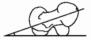
Femoral Neck Version


  • Also known as angle of femoral torsion, angle of antetorsion, angle of anteversion, angle of declination


image


Femoral neck version is the orientation of the femoral neck in relation to the femoral condyles at the level of the knee when the femur is viewed along the axis of the shaft. In most cases, the femoral neck is anterior to the transcondylar femoral axis (called anteversion). When the neck axis is posterior to the condylar axis, it is termed retroversion.

Birth 30°-40°

Adult: Males, 8°; females, 14° (Normal ranges are very broad.)


Genu Varum/Valgum

Genu varum is a frontal plane deformity in which the knee joints are angled to where they are close together (genu valgum), also called “knocked knee,” or where they are angled apart (genu varum).


image


Tibia


Tibial Torsion

Tibial torsion is the lateral twist of the long axis of the tibia. Tibial torsion is measured clinically by malleolar position, which is the angle between the knee axis and the medial and lateral malleoli. Note that the malleolar position is about 5° less than the tibial torsion angle.


Age


Tibial Torsion


Malleolar Position


Birth


1 y


3°-4°


2 y


12°


6°-8°


6 y


22°


13°-18°


Adults


18°-23° (ave 20°)


image


Varum/Valgum

A frontal plane deformity in which the long axis of the distal third of the tibial is angled either medially or laterally from the midsagittal plane of the more proximal two-thirds of the tibia. Tibial varum is compensated in the foot with subtalar joint pronation, and tibial valgum is compensated by supination. Tibial varum and valgum must be evaluated in conjunction with knee angles (genu varum and valgum). A
genu varum of 5° plus a tibial varum of 5° mean that the subtalar joint must pronate10° to allow the calcaneus to assume a vertical position.


Birth


5°-10° varum


>2 y


2°-3° varum (normal adult values)


image


Talocrural Angle

A line drawn parallel to the tibial plafond and a second line drawn connecting the tips of the medial and lateral malleoli. This measurement is useful in assessing syndesmotic injuries and ankle fractures. Normal value is 83° ± 4.


image

Only gold members can continue reading. Log In or Register to continue

Stay updated, free articles. Join our Telegram channel

Nov 20, 2018 | Posted by in ORTHOPEDIC | Comments Off on Biomechanics

Full access? Get Clinical Tree

Get Clinical Tree app for offline access