Fig. 5.1
Injury rate by traumatic mechanism (Data from a 3-year observational study on an Italian first league basketball academy)
Also playground, surfaces, and sports equipment influence the pattern of injury [14]. Competitions have a bigger injury ratio than practice, both in professional and amateur basketball categories [5, 15]. Related to training time, different authors showed that those who train longer have more injuries compared with those who train less. Probably the increased exposure may be related to an increased risk of injury due to repetitive and cumulative trauma [7, 16, 17]. Different data analysis observed that most basketball injuries resulted in at least 10 consecutive days of restricted or total loss of participation [6].
Position played by an athlete and anthropometric characteristics are other potential factors that influence the risk of injury. A center is most of the time under the basket in a high–player density area and is thus more at risk for contact injury than a player at the periphery, such as a point guard or forward [18, 19]. An analysis on Brazilian professional basketball players showed that the highest number of injuries concerned center players (44.1 %), followed by forwards (35.3 %) and guards (20.6 %). Center players suffered hand, chest, and abdomen trauma and sprained ankle more than others, mostly after he moves in the free-throw lane and has more physical contact to catch rebounds or for short shots. On the other hand, they have less nontraumatic injuries, probably because their movements are not as intense as forwards and guards, who presented a high rate of nontraumatic injuries [20].
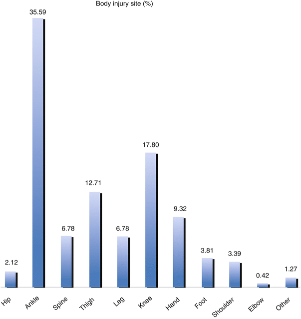
In basketball lower extremity injuries predominate; the ankle is the most frequently injured anatomic site and specifically ankle sprains representing the most common injury (Fig. 5.2) [6, 8].


Fig. 5.2
Injury rate by body site (Data from a 3-year observational study on an Italian first league basketball academy)
Injuries to the distal lower extremity have a multifactorial etiology; there is an interaction of psychological, physiological, biomechanical, and anthropometric factors. It was observed that the tissue composition of the leg, specifically the ratio of fat mass to bone mineral content, is related to distal lower extremity injury. The bone mineral content and stiffness of the lower extremity are also inversely correlated to the playground surface with no clear consequences in shock experience [21–23].
Knee and back injuries are also relatively prevalent, whereas injuries at the hip and groin occur less frequently [8]. In the upper extremity, hand and wrist injuries are most commonly encountered [5, 6].
Both in male and female, young and adult players, there are commonalities and differences in injury patterns [2, 3, 24, 25]. Some studies state that female athletes have increased risk for injury; however in literature there is no unambiguous consent regarding this statement [26–29].
Characteristics of children basketball players’ musculoskeletal system make them vulnerable to injuries unseen in adults. Open physis trauma could result in fractures, while in an adult we see strains and sprains because tendons, ligaments, and capsules are stronger than physeal cartilage. The physis is particularly vulnerable during times of rapid growth. The pull of a strong tendon near a growth center can result in repetitive traction injury, as occurs in the Osgood–Schlatter lesion. In a rapidly growing child, there could be differences in growth rates of bones and soft tissues which can result in a loss of flexibility and coordination and muscle imbalances [30–33]. Following gender comparisons of young basketball players, boys are more predisposed to lacerations, fractures, and dislocations, whereas girls suffer mainly traumatic brain injuries, sprains, strains, and soft tissue injuries, and frequently knees and upper extremities are involved. Randazzo et al. described in their data how upper extremity injuries (specifically to the finger) and traumatic brain injuries are more common in younger children (5–10 years of age), whereas lower extremity injuries (specifically to the ankle), sprains and strains, and lacerations are more frequent in older children and adolescents (15–19 years of age) [34].
5.1 Acute Injuries
As acute injury we define any basketball accident requiring minimum care and causing the absence of the injured player for at least one training or game session [35].
5.1.1 Ankle Sprains
Ankle sprains are the most common acute injuries in adult as well as young basketball players, both in professional players and amateur players. The risk of ankle sprains is higher during games than during training and more likely in offense than defense [3, 6, 8, 24, 36–38]. Lateral ankle sprains predominate over medial sprains, and some researchers refer to ankle sprain as exclusively an injury of the lateral ligament complex [39–41]. The most frequent traumatic mechanism in lateral sprains is an inversion injury with the foot in slight plantar flexion. This happens mostly when one player steps on the foot of another player rolling the ankle inwards or when the player lands awkwardly. Cutting, turning, and pushing off awkwardly are other common causes for ankle sprains. The anterior talofibular ligament is most commonly injured followed by the calcaneofibular ligament. Eversion injury is much less common, occurring secondary to either dorsiflexion with eversion or external rotation of the foot. Nevertheless, eversion injuries may be relatively serious because the deltoid ligament, anterior tibiofibular ligament, and interosseous membrane may be involved with the disruption of the ankle mortise [3, 39, 40]. Magnetic resonance imaging (MRI) is useful to confirm ligament deficiency and to detect injuries that are radiographically occult, including chondral or osteochondral injuries of the talar dome [40, 42–44].
Ankle taping and bracing, high-top shoes, and balance training show in different studies a protective effect on the rate of ankle sprains in basketball; they are particularly effective in players with prior ankle injuries. Thacker et al. found semirigid ankle braces to be effective in preventing ankle sprain and that braces do not adversely affect performance [13, 45–49]. A lot of ankle sprains sustained during basketball become recurrent, and a large part of these players perceived mechanical instability and persisting symptoms [50, 51].
It’s very important to evaluate ankle injuries and fractures in child athletes accurately due to the presence of open growth physes and make sure to exclude a Salter I fracture of the distal fibula, which may only be suggested on radiographs by soft tissue swelling adjacent to the injured growth plate [52].
A lot of acute foot injuries are associated to an ankle sprain. These injuries include avulsion fractures of the navicular, avulsion fractures of the base of the fifth-ray metatarsal (secondary to inversion stress), avulsion at the origin of the extensor digitorum brevis muscle from the lateral calcaneus, fractures of the anterolateral process of the calcaneus (typically occur with the foot abducted and plantar flexed), fractures of the anterolateral process of the calcaneus, os trigonum fractures, and tear of the superior peroneal retinaculum (SPR) with peroneal tendon subluxation (it may occur during acute dorsiflexion with strong contraction of the peroneal muscles to prevent further dorsiflexion) [42].
5.1.2 Knee Ligament Acute Injuries
The mechanism of injury to the anterior cruciate ligament (ACL) in basketball is more commonly noncontact, deceleration and sudden change in direction that may cause abnormal rotation of the tibia, resulting in ACL injury. Furthermore valgus knee collapse occurs more frequently in women [53, 54].
Different studies show a strong female predilection in ACL injury in basketball players; different meta-analysis of the incidence of ACL injury as a function of gender and sport reveal a female to male ratio of 3.5:1 [7, 25, 55, 56]. The following risk factors can explain this increased risk of ACL injury in female adult and young athletes: a heavy risk in the preovulatory phase of the menstrual cycle, decreased intercondylar notch width on radiographs (this factor is not confirmed in professional male basketball players), and a predisposition to increased knee abduction on landing in female athletes [33, 57, 58].
A common pattern of ACL injury for a skeletally immature athlete is an avulsion fracture of the tibia, or less commonly the femur, at the site of ACL attachment, as the chondro-osseous junction is the weakest part of the ACL complex [33].
ACL tear is a serious injury with significant loss of playing time and long rehabilitation after surgical repair. Although many competitive basketball players return to action after ACL reconstruction, Busfield et al. showed that 22 % of NBA players didn’t return to compete and 44 % of those who returned experienced a lower player efficiency rating [59].
Injuries to the posterior cruciate ligament (PCL) are extremely rare in basketball; the mechanism in an athlete typically involves a fall on the shin or a hyperextension injury [60].
Avulsion of the tibial tubercle in a young athlete has been well described among basketball players and may occur bilaterally. The mechanism involves violent knee flexion against a tightly contracted quadriceps muscle or violent quadriceps contraction with a fixed foot [33].
5.1.3 Hip and Pelvis Acute Injuries
Compared to ankle and knee injuries, injuries to the pelvis, hip, and upper thigh are moderately common in basketball players. According to the National Collegiate Athletic Association (NCAA) and NBA, injuries to the pelvis, hip, and upper leg accounted for approximately 10 % of game-related injuries and 11 % of injuries sustained during practice. In all data thigh injuries are more prevalent, and the specific injuries identified were musculotendinous strains and contusions [2, 6, 41]. Although there is a heightened understanding of intra-articular hip pathology, most athletic-related injuries to the hip are extra-articular [10]. Most injuries occurring about the pelvis, hips, and upper thighs were composed of musculotendinous strains and contusions, which included adductor and rectus abdominis strains, hamstring injuries, and thigh muscle contusions. Hamstring and adductor tears have been shown to be predominately proximal and constitute injury about the hip rather than the knee [61, 62]. The quadriceps is the most commonly injured (contusion/strain) structure and has significant game-related injury rate compared with other structures. The hamstring muscle group was the most frequently strained as result from dynamic overload/eccentric contractions. Jackson et al. demonstrated in their statistical analyses that strains were most frequent in the first month of the season (the preseason) and the cumulative risk is related with length of the player career and of each season [10].
5.1.4 Upper Extremity Acute Injuries
Upper extremity injuries, overall, accounted for 12–13 % of injuries sustained at both the high school and professional levels of play [5, 8]. Hand and arm injuries predominate over injuries to the shoulder or elbow [63]. The fingers and thumb represent the most likely site of acute fracture in basketball players, and the proximal interphalangeal joints (PIP) are the most frequently injured sites [7]. “Dunk lacerations” have also been described in basketball players. These injuries occur secondary to the impact of the player’s hand with sharp edges of the rim or with the flange connecting the rim to the backboard [64]. A mallet finger, also known as a hammer finger, is a very common basketball injury that disrupts the extensor mechanism at the distal interphalangeal joint. The mechanism is typically an axial load on a partially flexed finger, usually a ball striking the tip of the finger. In a young athlete, an avulsion fracture of the epiphysis is more likely than a rupture of the extensor tendon. Other hand/finger injuries may also include tears of the volar plate (sometimes associated with an avulsion fracture), metacarpophalangeal joint injuries (thumb’s ulnar collateral ligament tear is very common), and carpometacarpal joint injuries [65, 66]. It’s an accepted opinion that for young children, age-appropriate basketballs should be used, which may decrease the rates of concussions and finger-related injuries, and rough play should be discouraged, to minimize collisions [34].
Shoulder injuries sustained during basketball are uncommon. In the NBA, the most frequently identified shoulder injuries were glenohumeral sprain, acromioclavicular joint sprain, and rotator cuff inflammation. In a review of shoulder injuries in high school athletes between 2005 and 2007, the incidence was 0.47 injuries per 10,000 exposures for boys and 0.45 injuries per 10,000 exposures for girls. Injuries were much more commonly sustained during competition than during practice, with most injuries occurring during defending and rebounding [8, 67].
5.1.5 Wrist Acute Injuries
Wrist injuries are most typically the consequence of falling on an outstretched hand. The location of a fracture following this type of fall depends on the angle of the wrist upon hitting the ground as well as the age of the patient at the time of injury. If the wrist is more flexed, the athlete is more likely to sustain a scaphoid fracture. If the wrist is more extended, he or she is more likely to suffer a distal radial or ulnar fracture. Also, the risk of scaphoid fracture in children increases as the bone matures [68, 69].
Ligamentous wrist injuries can include injuries to the triangular fibrocartilage complex (TFCC). This type of injury may occur due to acute trauma or repetitive injury. When acutely injured, the mechanism is often related to axial load bearing with rotational stress, often during a fall on an outstretched hand [68].
5.1.6 Back Acute Injuries
Back injuries in basketball players accounted for 6.8 % of all injuries sustained by NBA players over a 10-year period but represented 11 % of all days missed. Back muscle strain was the most common presentation; disk rupture/herniation was far less prevalent. Cervical spine injuries were significantly less common than lumbar injuries, accounting for 1.3 % of injuries overall. Sacral injuries amounted to 0.6 % of the total and thoracic spine injuries 0.5 % of the total [8].
5.2 Overuse Injuries
As overuse injuries we define those causing physical discomfort with an insidious onset; they cause pain and/or stiffness and can potentially affect the player during and/or after the basketball activity [3].
5.2.1 Ankle, Foot, and Lower Leg Overuse Injuries
MRI is considered the gold standard for early diagnosis of stress injury. Osseous stress fractures occurring below the knee in the basketball player frequently involve the tibia or the distal fibula, the latter most common approximately 5 cm proximal to the tip of the lateral malleolus. Stress fractures may also be observed, albeit less commonly, in the tarsal navicular, calcaneus, metatarsals, and cuneiforms [70].
Tibial stress fractures most commonly involve the posteromedial cortex, a pattern of injury most commonly seen in athletes participating in running sports. Athletes involved in jumping sports such as basketball can develop a more specific stress fracture involving the anterior tibial cortex. For stress fractures of the posteromedial surface, the prognosis is generally good with conservative management. The prognosis is worse if the fracture involves the anterior tibia, as these have a higher rate of nonunion or progression to complete fracture [71, 72]. Potentially modifiable risk factors for stress fractures in female basketball players include low cardiorespiratory fitness, lack of resistance training, poor nutrition (e.g., low calcium intake, negative energy balance), menstrual dysfunction, shoes, and less time to recover with hard off-season workouts [73–75].
One severe foot injury in basketball players, which may result from overuse, is the Jones fracture. The Jones fracture is located at the diaphyseal-metaphyseal junction of the proximal fifth metatarsal and results from the abnormal loading of the lateral foot when the heel is elevated and the metatarsophalangeal (MTP) joints are hyperextended [76]. Jones fractures may be slow to heal, and they have a high rate of nonunion when treated nonoperatively [77].
Several soft tissue conditions in the lower leg and foot of basketball players may result from overuse. Achilles tendinosis may be both insertional and non-insertional in location. At the ankle and hindfoot, plantar fasciitis is not uncommon, and anterior ankle impingement may be a sequel of chronic lateral ankle sprains with ligament injury.
At the forefoot, sesamoid and MTP joint injuries, synovitis, and adventitial bursa formation may be observed [42, 76]. Sports-related Achilles tendon rupture peaks in the fourth decade. Degenerative change is usually present in complete rupture, and most ruptures occur 2–6 cm above the calcaneus, where the blood supply is lowest, and flow decreases with age [78–80]. Achilles tendon injuries occur when significant forces are translated through a malaligned tendon with a dorsiflexed foot and an extended knee. Of the 18 players identified by Amin et al. over 23 NBA seasons, only 44 % were able to return to play for longer than 1 season after their surgical repair. Those who did return to play did not perform as well as their control-matched peers [81].
5.2.2 Knee Overuse Injuries
Overuse injuries at the knee predominate at the extensor apparatus. Some investigators consider the entity jumper’s knee to apply to tendinosis occurring anywhere along the extensor mechanism from quadriceps tendon to the tibial tubercle and not exclusively to that involving the proximal patellar tendon [82]. In a 10-year prospective study of injury and illness in the NBA, patellofemoral inflammation accounted for 8.1 % of orthopedic injuries [8].
Eccentric muscle contraction inherent in sudden decelerations and jumping can result in extensor mechanism injuries, including the patellofemoral joint. These mechanisms may lead to microscopic tendon tear, especially in the proximal patellar tendon. Similarly, excessive force generated across the patellofemoral articulation may result in overuse injury, especially in people with underlying joint malalignment. Heavy and long-standing abnormal stress may even lead to patellar or quadriceps tendon rupture or patellar stress fractures [82]. Early patellofemoral arthrosis in basketball players may be asymptomatic. Whether or not symptoms are present, patellofemoral chondromalacia may manifest as cartilage signal alteration, fissuring, or fibrillation. Focal cartilage defects along patella and trochlea may be observed, often with underlying subchondral cysts or bone marrow edema [83].
Stay updated, free articles. Join our Telegram channel

Full access? Get Clinical Tree


