51 Axial skeleton trauma
The axial skeleton comprises the skull (see Ch. 72) and spine, and it is also useful to consider the pelvis here. Trauma victims are always initially assessed by ABC principles including immobilization of the cervical spine and treatment of shock.
 SPINAL TRAUMA AND VERTEBRAL FRACTURES
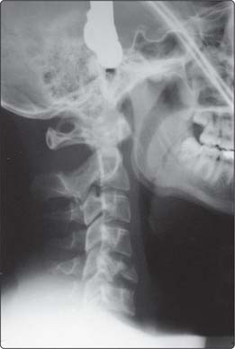
Most spinal injuries occur in accidents, primarily RTAs (50%), accidents in the home and workplace, or sports injuries. Cord injury is only seen in 5%. All unconscious patients should be assumed to have a spinal injury; a fractured vertebra is unstable until proved otherwise. A urinary catheter can be passed since the patient may lose filling reflexes with lower spinal injuries and go into painless urinary retention. Clearing the cervical spine is done with a combination of clinical, neurological and radiographical examinations. Radiographs include a lateral radiograph to show the first cervical to first thoracic vertebrae (C1 to C7/T1 junction; if it does not do this it is deemed inadequate) (Fig.3.51.1) and A-P views of the cervical spine and through the open mouth to show the odontoid peg (peg view). The radiographs are assessed using the ABCS system:
 alignment of vertical contours formed from the vertebral bodies, spinal canal and spinous processes: a step of > 25% suggests a unifacet joint dislocation, and > 50% suggests a bifacet dislocation
 bone margin and density: outline of vertebral bodies and spinous processes for avulsion and wedge fractures
Stay updated, free articles. Join our Telegram channel

Full access? Get Clinical Tree


