Athlete with Syncope


Athlete with Syncope


Christopher Lutrzykowski


INTRODUCTION

Syncope is defined as the temporary loss of consciousness and posture. Near syncope is defined as a prodromal symptom of fainting or near faint. These events can occur before, during, or after an athletic event and warrant an investigation. A cause for syncope can be found in up to 50% of patients.1 A good patient history may be the single best determinant in guiding the clinician to the correct diagnosis.1, 2, 3 The history is often obtained by interviewing the athlete, but it is paramount to include those who witnessed the event. This group includes parents, coaches, and trainers who can be of incalculable value in narrowing the differential diagnosis. Using the appropriate screening questions in a preparticipation examination may also be of benefit, as shown in Table 3.1.2,3

The athlete with syncope presents a diagnostic challenge due to the large list of potential etiologies ranging from the benign to the catastrophic. An excellent resource for the clinician caring for athletes is the 36th Bethesda Conference report: Eligibility Recommendations for Competitive Athletes with Cardiovascular Abnormalities.

There continues to be a highly visible and defined group of athletes who suffer a sudden cardiac death (SCD) event. This chapter includes guidelines for caring for the athlete with a variety of cardiac conditions including hypertension, arrhythmia, and syncope.


PATHOPHYSIOLOGY

Syncope occurs secondary to a sudden loss of blood pressure (BP) to the brain. Etiologies in athletes include both cardiac and noncardiac types. Hypertrophic cardiomyopathy (HCM) is a relatively common inherited medical condition, with the incidence estimated at 1:500 in the general population, and is inherited in an autosomal dominant fashion.5 It is caused by mutations in any one of 10 genes that encode proteins of the cardiac sarcomere.6 Initially thought to be a disease of white males, recent studies indicate a proportionate incidence among many ethnic groups.6,7 Indeed it is estimated to be underdiagnosed in these populations as well as among women.7,8 Sudden death related to HCM also crosses ethnic lines with similar prevalence rates on autopsy of athletes after SCD.8

Postexertional syncope, also known as exercise-associated collapse, is a benign condition caused by venous pooling upon the completion of exercise. The cessation of normal muscular contraction of the lower extremity results in transient decreased venous return and subsequent cerebral hypoperfusion. Arrhythmias can occur for a variety of etiologies, with ischemia being the leading cause in the athlete over the age of 35. A full description of arrhythmias is listed later in the chapter.


EPIDEMIOLOGY

The rate of syncope among athletes is not known, but cardiac causes appear more defined. A recent study of 7,500 athletes revealed a syncopal history in 6% of athletes over a 5-year period.9 This same study also noted that only 1% of those athletes had exertion-related syncope and no postexercise or nonexercise syncopal event was associated with structural heart disease.9 The incidence of SCD has been estimated to be 1 per 200,000 athletes yearly in the United States, but true incidence may be higher.10,11 The rate among young adults is less than 1% than that of SCDs among adults with the total number of cardiac-caused events reaching 10 to 13 per year.1,12 Most causes of syncope are benign but deserve a workup due to both the potential for SCD as well as the disruptive nature of the event.13 The majority of SCDs occur during sports activity or the immediate period thereafter. However, 20% of SCDs may occur in the ensuing 24-hour period post exercise, further complicating the correct identification of the true incidence rate.14,15 Syncope can be recurrent; noncardiac syncope has a recurrence rate approaching 43% at 5 years.16 The most common cause of nonexertional syncope among young competitive athletes appears to be vasovagal or “situational” (88% and 12%, respectively). Postexertional syncope (exercise-associated collapse) is most commonly associated with postural hypotension. Exertional syncope is most commonly associated with exertional-induced neurally mediated
syncope (66%). Cardiac anomalies appear to account for the rest.9 Sudden death due to cardiac causes is most often attributed to HCM; as much as 50% of cardiovascular SCDs may be attributable to HCM.15,17 HCM is followed by coronary artery anomalies, left ventricular hypertrophy (LVH), myocarditis, Marfan syndrome, and arrhythmogenic right ventricular cardiomyopathy in order of prevalence.4,6,17 Latent coronary artery disease (CAD) dominates the incidence of SCD after the age of 35 with vast majority of causes (80%).17 Table 3.2 lists the causes of SCD based on order of frequency.


TABLE 3.1 AHA Consensus Panel Recommendations for Preparticipation Athletic Screening4


Family History


Personal History


Physical Examination


Premature sudden cardiac death


Heart murmur


Heart murmur (supine/standinga)


Heart disease in surviving relatives younger than 50 years old


Systemic hypertension


Femoral arterial pulses (to exclude coarctation of aorta)


Fatigue


Syncope/near syncope


Stigmata of Marfan syndrome


Excessive/unexplained exertional dyspnea


Brachial blood pressure measurement (sitting)


Exertional chest pain


a In particular, to identify heart murmur consistent with dynamic obstruction to left ventricular outflow. Modified from Maron BJ, Thompson PD, Puffer JC, et al. Cardiovascular preparticipation screening of competitive athletes. A statement for health professionals from the Sudden Death Committee (clinical cardiology) and Congenital Cardiac Defects Committee (cardiovascular disease in the young), American Heart Association. Circulation. 1996;94:850-856.


TABLE 3.2 Causes of Sudden Death in 387 Young Athletes


Cause


No. of Athletes


Percent


Hypertrophic cardiomyopathy (HCM)


102


26.4


Commotio cordis


77


19.9


Coronary artery anomalies


53


13.7


Left ventricular hypertrophy of indeterminate causationa


29


7.5


Myocarditis


20


5.2


Ruptured aortic aneurysm (Marfan syndrome)


12


3.1


Arrhythmogenic right ventricular


11


2.8


Tunneled (bridged) coronary arteryb


11


2.8


Aortic valve stenosis


10


2.6


Atherosclerotic coronary artery disease


10


2.6


Dilated cardiomyopathy


9


2.3


Myxomatous mitral valve degeneration


9


2.3


Asthma (or other pulmonary condition)


8


2.1


Heat stroke


6


1.6


Drug abuse


4


1.0


Other cardiovascular cause


4


1.0


Long QT syndromec


3


0.8


Cardiac sarcoidosis


3


0.8


Trauma causing structural cardiac injury


3


0.8


Ruptured cerebral artery


3


0.8


a Findings at autopsy were suggestive of HCM but were insufficient to be diagnostic.

b Tunneled coronary artery was deemed the cause of death in the absence of any other cardiac abnormality.

c The long QT syndrome was documented on clinical evaluation. Modified from Maron BJ. Sudden death in young athletes. N Engl J Med. 2003;349:1064-1075.


Adapted from data from the registry of the Minneapolis Heart Institute Foundation.14


The Athlete’s Heart

The athlete’s heart undergoes physiologic changes with exercise, which impact diagnostic and therapeutic considerations. Cardiac remodeling and increased ventricular mass occur as a normal adaptation to prolonged exercise. In addition,
atrial and ventricular cavity size enlargement, without systolic or diastolic dysfunction, is also present because of increased blood volume. This is defined as the athlete’s heart. These changes may occur within weeks of beginning a training program, are probably proportional to the intensity and duration of training, and resolve with decreasing exercise loads.18 Electrocardiogram (ECG) findings in athletes can mimic cardiac injury patterns and may lead to unnecessary exclusion from participation.4 Conversely, adolescent athletes rarely exhibit deep T-wave inversions;19 2% of athletes exhibit increased left ventricular (LV) wall thickness of 13 to 15 mm, thus overlapping the diagnostic criteria for HCM; and 15% of athletes have LV chamber size greater than 60 mm, which can raise concern for cardiomyopathy despite normal LV function.20 Referral for further diagnostic testing is warranted, including echocardiogram (ECHO) (Fig. 3.1). Careful consideration should be given to athletes in this gray zone, as the risk for SCD is considerable, yet overly strict guidelines may exclude many athletes not otherwise at risk. ECG findings in athletes may return to normal, but ECHO findings continue to display HCM in those athletes with the disease.21 Adolescent athletes rarely exhibit chamber size greater than 12 mm; LV wall thickness greater than 12 mm should prompt further evaluation for HCM.22 Athletes with LV wall thickness greater than 15 mm on ECHO should be referred to a cardiologist.


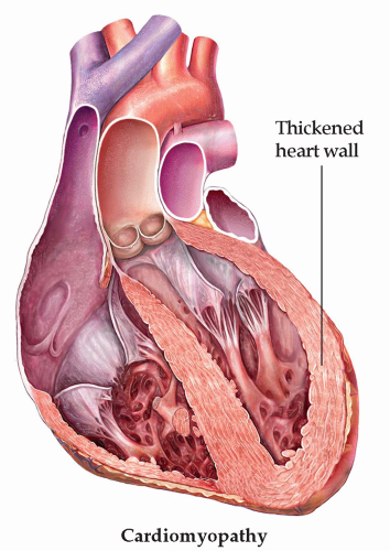
FIG. 3.1. Left ventricular hypertrophy. Modified from Opie LH. Heart Physiology: From Cell to Circulation. Philadelphia: Lippincott Williams & Wilkins; 2004. © LH Opie, 2004.


NARROWING THE DIFFERENTIAL DIAGNOSIS


History

An adequate history of the event must be obtained from the athlete, coaches, parents, and trainers. It is a necessary but often time-consuming process. The history will often lead to the diagnosis and guide further workup and should include specific details of the event. The timing of the syncopal event should be noted; did the event occur before, during, or after the competition? How long after the event? Any prodromal symptoms such as palpitations or chest pain should be noted. Prior history and family history should be noted, including family history of sudden death. A dietary history should be obtained, including intrarace nutrition. Any medications, supplements, or ergogenic aids should be discussed and explored. The overall duration of the event and specific interventions should also be noted. Any physical stigmata of note, such as seizure activity, should also be recorded. A collapse “mid stride” is ominous as the majority of SCDs in athletes occur during or just after athletic participation.6,21

Only gold members can continue reading. Log In or Register to continue

Stay updated, free articles. Join our Telegram channel

Sep 22, 2016 | Posted by in ORTHOPEDIC | Comments Off on Athlete with Syncope

Full access? Get Clinical Tree

Get Clinical Tree app for offline access