Fig. 5.1
DXA scan of the lumbar spine of a healthy 14-year-old girl. Areal BMD (aBMD) is calculated from BMC by dividing BMC by the projected area in the coronal plane of the region scanned. Despite a low T-score, the Z-score is normal, implying a normal bone density. In athletes under the age of 20 years, Z-scores should be used instead of T-scores. Images were obtained with a Hologic QDR-4500 scanner
Based on data in postmenopausal women, a 1 standard deviation (SD) reduction in aBMD is associated with a twofold increase in fracture risk and in adults, osteoporosis has been operationally defined as a BMD ≤ 2.5 SD below the young adult mean (T-score ≤ −2.5) and osteopenia as a T-score < −1.0. In children and adolescents, Z-scores (the number of SD below the age-matched mean) should be used instead of T-scores. A low BMD T-score in a 14-year-old athlete who has not yet achieved peak bone mass may be perfectly normal when compared with age-matched controls (Fig. 5.1). In addition, because DXA measures aBMD rather than volumetric density (vBMD, expressed as g/cm3), it can underestimate true volumetric density in small children and adolescents and overestimate it in larger children and adolescents. Furthermore, some female athletes (such a gymnasts and long distance runners) may have short stature or pubertal delay. Correction methods are now available that adjust for height that can improve the accuracy of the BMD Z-score [19–21].
Unlike studies in adults in whom a T-score in the osteoporotic range is associated with an increased fracture rate, in children and adolescents, there is no specific Z-score below which fractures are more likely to occur, but there is a growing body of evidence demonstrating an association between low aBMD and increased fracture risk [22–24]. The International Society for Clinical Densitometry (ISCD) recommends that in males and females younger than 20 years of age, the term “osteopenia” should no longer be used and the term “osteoporosis” should not be based on bone densitometry results alone. The ISCD defines low BMD for chronological age as an age, sex, and body size-adjusted BMD Z-score < −2 [14, 25]. The ISCD further recommends that in those under 20 years of age, the diagnosis of “osteoporosis” requires both a low BMD or BMC for age (Z-score < −2) plus a clinically significant fracture, defined as a long bone fracture of the lower extremity, a vertebral compression fracture, or two or more long bone fractures of the upper extremity [25]. Fractures of digits or toes are not included in this definition of a “clinically significant fracture” [26]. Similarly, a child or adolescent with low BMD for chronological age but without a clinically significant fracture does not meet criteria for osteoporosis.
Because bone mass is expected to be higher in athletes than in sedentary individuals, the ACSM recommends that in athletes, a BMD Z-score less than −1.0 warrants further investigation, even in the absence of a prior fracture [3]. A recent prospective cohort study of 163 female high school athletes, revealed that a BMD Z-score < −1.0 was associated with a 3.6-fold increased risk of musculoskeletal injury (not necessarily fracture) during the interscholastic sports season compared with athletes who had normal BMD values [27]. These findings support the ACSM’s recommendations.
Bending strength of a bone depends not only on BMD, but also on bone elasticity and bone geometry. Section modulus, an engineering term used to estimate bending strength of a hollow structure, can be calculated from DXA scans using Hip Structural Analysis, an interactive computer-based program that calculates section modulus from measurements of the cross-sectional area of the femoral neck using images derived from the DXA scan [28]. Section modulus of a large bone will always be greater than that of a smaller bone, even when both bones have the same BMC or BMD. In adults, assessment of bone geometry based on DXA scans is predictive of hip fracture risk, independent of bone density [29, 30]. In young girls, Hip Structural Analysis has been used to demonstrate the positive effects of jumping activities on section modulus of the femoral neck in early pubertal girls [31].
When to Order DXA Scans
There is no strong evidence to guide clinicians when to order DXA scans and a decision to do so for an individual patient still requires clinical judgment. Consensus of expert opinion recommends that DXA scans should be considered in a female athlete with recurrent fractures, a “clinically significant” fracture (defined as a fracture of a long bone of the lower extremity, a vertebral compression fracture, or two or more fractures of long bones of the upper extremity), a low impact fracture (defined as a fracture sustained from standing height or less), or in an athlete who has been amenorrheic for more than 6 months [26, 32]. Repeat DXA scans should be performed at an interval that can identify a change between the two DXA assessments that exceeds the error of repeated measurements. Based on expert opinion, the ISCD recommends a minimal interval of 6 months before repeating scans [25]. A recent study demonstrated that precision error of DXA scans varies with region of interest, age, and sex. For girls 17 years and younger, a monitoring time interval of 1 year enabled identification of DXA changes that exceeded precision error [33]. Until further information becomes available, for most adolescents and young adults, it is reasonable to repeat the DXA measures after 1 year. The same machine should be used for serial scans in order to make an accurate assessment of the percent change in BMD over the prior year.
Quantitative Computed Tomography
Quantitative computed tomography (QCT) is a three-dimensional imaging modality that accurately measures true vBMD and can differentiate cortical bone from trabecular bone. QTC measurements of the spine and hip are obtained using a clinical whole body scanner that is equipped with special analysis software. Bone size and geometry can be assessed and the scanner can also be used to measure bone density at the distal forearm. QCT machines are costly, not readily available, and utilize high doses of radiation (30–7,000 μSv) [34].
Newer modalities such as peripheral QCT (pQCT) can measure vBMD of the appendicular skeleton with much lower doses of radiation (<3 μSv). The pQCT machines are smaller and more mobile than a clinical whole body scanner and are dedicated to assessment of bone health. Usual sites measured are the non-dominant distal tibia and distal radius. The use of pQCT is particularly appealing for assessment of bone health in athletes because an athlete is more likely to sustain a fracture of the arm or leg than of the hip or spine. In addition to measurement of vBMD, pQCT can measure cortical thickness, cortical density, and trabecular density from cross-sectional images generated. In a cross-sectional study of 204 competitive female athletes using pQCT, Nikander et al. demonstrated that, compared to athletes participating in low impact sports or those in a control group, athletes in high-impact sports had enhanced bone geometry of the tibia evidenced by a thicker cortex at the distal tibia and a greater cross-sectional area of the tibial shaft [35]. A study of 396 Finnish girls aged 10–13 years using pQCT showed that girls who sustained upper limb fractures during puberty had low vBMD of the distal radius at age 10–13 that persisted into adulthood, confirming prior DXA studies regarding the relationship between BMD and fracture risk in children [36].
High resolution pQCT (HR-pQCT) measures small regions of the distal tibia and radius and can evaluate bone microstructure (cortical thickness, trabecular number, thickness, and separation), as seen in Fig. 5.2. It can also be used to estimate bone strength. Scanning time is <3 min and dose of radiation is low (<3 μSv). In postmenopausal women, use of HR-pQCT was better able to predict fragility fractures than measures of BMD performed by DXA [37]. In adolescents, HR-pQCT has been successfully used to assess bone microstructure while avoiding irradiation of the active growth plate [38]. Finite element analysis, FEA, is a computer-based modeling technique used to reconstruct three-dimensional images of the bone in order to estimate bone strength by calculating the predicted load necessary to fracture the bone. Studies using HR-pQCT-based FEA have shown that estimation of bone strength using FEA can enhance prediction of wrist fractures in postmenopausal women [39]. Use of HR-pQCT, while still limited to research, shows great promise for clinical use and has the potential to better predict fracture risk than DXA. Unfortunately HR-pQCT machines are only found in a select few bone research centers. A summary of the advantages and limitations of different imaging modalities is shown in Table 5.1.
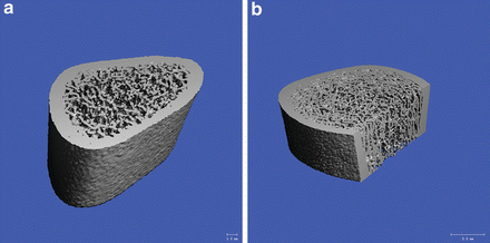
Fig. 5.2
HR-pQCT images of the distal radius (a) and distal tibia (b) of a healthy 14-year-old girl, demonstrating the trabecular microarchitecture and cortical shell. Images were measured at the 7 % site for the radius and at the 8 % site for the tibia using an Xtreme CT (HR-pQCT) scanner. [Images courtesy of Dr. Melissa Putman and Dr. Catherine Gordon]
Table 5.1
Advantages and limitations of different imaging modalities
DXA | QCT | pQCT | HR-pQCT | |
|---|---|---|---|---|
Site measured | Lumbar spine | Lumbar spine | Distal radius | Distal radius |
Hip | Hip | Distal tibia | Distal tibia | |
Total body | Distal radius | |||
Radiation dose (μSv) | 5–6 | 30–7,000 | <3 | <3 |
BMD | aBMD | vBMD | vBMD | vBMD |
Differentiates cortical from trabecular bone | No | Yes | Yes | Yes |
Bone geometry | No | Yes | Yes | Yes |
Bone microstructure | No | No | No | Yes |
Magnetic Resonance Imaging
Magnetic resonance imaging (MRI) is a very sensitive method for detecting early stress changes in bone (including stress fractures) and can also provide detailed information about soft tissue injuries (see Chap. 4) [40]. There is no exposure to ionizing radiation but MRI scans are more expensive than DXA scans. MRI is not routinely used as a method of assessment of bone health but is frequently used to evaluate injuries.
Quantitative Ultrasound
Quantitative ultrasound is a noninvasive method of assessing bone health by measuring speed of sound of an ultrasound wave as it is propagated along the surface of bone. Ultrasound measures can be obtained on the calcaneus, tibia, and radius. The machine is easily portable and the test is relatively inexpensive and does not utilize radiation. However, initial enthusiasm for this method has been tempered by poor reliability of measurements, lack of pediatric reference databases, and uncertainty about what skeletal properties are captured by this assessment tool.
Biochemical Markers of Bone Metabolism
Measurement of markers of bone formation and degradation offers an opportunity to assess dynamic changes in bone turnover before these changes become apparent using traditional methods of assessment of BMD or BMC. Osteocalcin (OC) and bone-specific alkaline phosphatase (BSAP) are serum markers of bone formation that are released at different stages of osteoblast proliferation and differentiation. Since OC is incorporated into the bone matrix and is later released into the circulation during bone resorption, it can also be considered a marker of bone turnover. Commonly used measures of bone resorption are Type I collagen C-terminal telopeptide (ICTP), cross-linked C-telopeptide (CTX), and cross-linked N-telopeptide (NTX), which are measured in the serum or urine (Table 5.2). Levels of these markers vary with age and pubertal development. Pediatric reference data are now available for OC, BSAP, CTX, NTX, and ICTP [41]. Bone markers should not be used as a single assessment of bone health, but are best used to monitor dynamic changes over relatively short periods of time, for example for monitoring the response to antiresorptive therapy. Measurement of bone markers is still primarily used in research settings.
Table 5.2


Biochemical markers of bone metabolism
Related posts:


Stay updated, free articles. Join our Telegram channel
Full access? Get Clinical Tree
