Arthroscopy of the Ankle
Jorge I. Acevedo
Peter G. Mangone
DEFINITION
Arthroscopy of the ankle has become an invaluable tool for evaluating and treating pathology in the ankle joint.
Arthroscopy allows a minimally invasive approach to the structures of the ankle with a magnified view.
Detailed knowledge of the anatomy surrounding the ankle joint as well as the different structural variations is key to avoiding complications.
ANATOMY
The anteromedial portal is located medial to the tibialis anterior tendon at the level of the ankle joint (FIG 1). Care should be taken to avoid injury to the long saphenous vein and nerve usually located medial to the portal.
The anterolateral portal lies on the anterior joint line just lateral to the peroneus tertius tendon or alternatively lateral to the extensor digitorum longus tendons. The intermediate cutaneous branch of the superficial peroneal nerve lies in close proximity to this portal.
Posteromedial and posterolateral coaxial portals lie parallel to the bimalleolar axis (FIG 2A).

FIG 1 • Anatomic landmarks for anterior ankle arthroscopy. At the joint line, the anteromedial portal is made immediately medial to the tibialis anterior tendon and the anterolateral portal is created lateral to the extensor digitorum longus tendon.
The posterolateral coaxial portal (FIG 2B) is located immediately posterior to the peroneus longus tendon, and the posteromedial coaxial portal (FIG 2C) ideally lies between the posterior colliculus (of the medial malleolus) and the posterior tibial tendon. (Placement between the flexor digitorum longus and the posterior tibial tendon is also acceptable.)
The sural nerve is located an average of 6.6 mm from this posterolateral portal, whereas the posterior tibial nerve is found an average of 5.7 mm from the posteromedial portal.
For advanced arthroscopic ligament reconstruction, an anatomic safe zone exist between the intermediate branch of the superficial peroneal nerve and the sural nerve.
DIFFERENTIAL DIAGNOSIS
Anterior ankle impingement
Ankle arthritis or frozen ankle
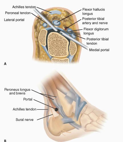
FIG 2 • Coaxial portal anatomy: cross-sectional (A), posterolateral (B), (continued)
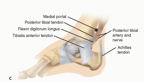
FIG 2 • (continued) and posteromedial (C).
Osteochondral tibial or talar defects
Lateral ankle instability
Ankle fractures
Recalcitrant ankle synovitis (often seen in patients with systemic inflammatory disease)
NONOPERATIVE MANAGEMENT
In general, conservative treatment will include a trial with activity modification, immobilization with a brace, and nonsteroidal anti-inflammatories.
Physical therapy using modality treatment, range-of-motion exercises, neuromuscular coordination training (eg, balance board), and strengthening of the secondary or dynamic stabilizing muscles surrounding the ankle is a useful adjunct to most conditions.
SURGICAL MANAGEMENT
Preoperative Planning
Imaging studies are reviewed to determine ideal portals to be used.
Standard anteromedial and anterolateral portals are sufficient to access the anterior and central tibiotalar pathology.
Posterior portals are considered when drilling posterior talar lesions or when it is necessary to address pathology (eg, synovitis, loose bodies) within the posterior capsule.
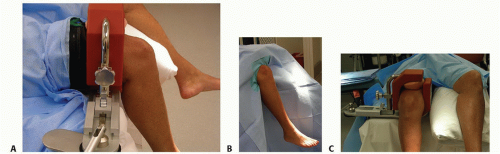
FIG 3 • A,B. Leg holder and bed positions, respectively, for posterior portal access. C. Position of operative leg and padded contralateral limb.
A preoperative popliteal block may be placed by anesthesia. Over the past 10 years, we have been able to perform 75% of ankle arthroscopies with regional anesthesia and light sedation.
An examination under anesthesia including anterior drawer as well as a talar tilt test should be performed before positioning.
Positioning
The patient is placed on a regular operating table with a well-padded tourniquet on the proximal thigh.
The supine position with a towel roll placed underneath the ankle is used when only anterior portals are necessary. In this situation, the tourniquet may be placed on the proximal calf.
If access to posterior portals is likely, then we lower the leg extension of the bed and use a standard arthroscopy knee holder (FIG 3A). This restricts thigh motion but allows free leg motion and access to the posterior hindfoot (FIG 3B). The contralateral leg is placed in a well-padded holder or pillow (FIG 3C).
Alternatively, a noninvasive ankle distractor is used.
Approach
The standard working approaches are the anteromedial and anterolateral portals.
Auxiliary anterior portals (such as the anterocentral) should be used with caution because of the high incidence of neurovascular injury.
The standard posteromedial and posterolateral portals should also be used with extreme caution due to the close proximity of neurovascular structures (FIG 4).
We prefer to use posterior coaxial portals parallel to the bimalleolar axis when addressing the posterior ankle joint.
Although the standard 4-mm arthroscope may be used, we prefer to use 2.7-mm arthroscopic instruments, which facilitate access and simplify the approach.
Instruments usually include 2.5-mm shaver, 3.5-mm shaver, thermal ablation device (this is especially helpful for synovectomy and débridement of the joint; however, care must be taken to avoid articular cartilage damage), and small arthroscopic biter and grabber devices.
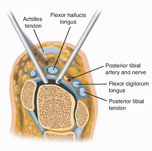 FIG 4 • Conventional posterior portal crosssectional anatomy.
Related posts:Stay updated, free articles. Join our Telegram channel
Full access? Get Clinical Tree
 Get Clinical Tree app for offline access

|
