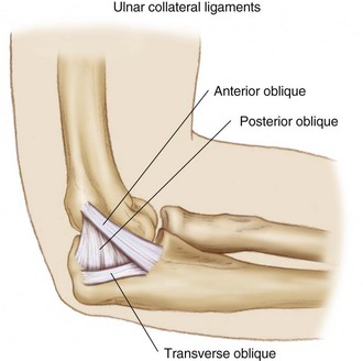
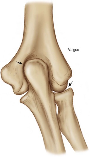
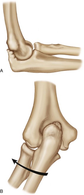
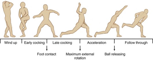
Chapter 41 The throwing motion in overhead athletes, particularly pitchers, places a tremendous amount of force across the medial ulnar collateral ligament (MUCL) on the medial side of the elbow. The anterior band of the MUCL is the primary restraint to valgus stress, especially at 20 to 120 degrees of flexion, where most athletic activity occurs (Fig. 41-1). The stress of the throwing motion can lead to developmental anatomic changes in the young thrower and places the overhead athlete at risk for specific injury patterns. Continued insult can lead to significant ligament and tendon injuries in the experienced athlete, valgus instability and attenuation of the MUCL, and posterior osteophyte formation. Figure 41-1 The medial ulnar collateral ligament is composed of the anterior band, posterior band, and transverse band. During the pitching cycle the valgus stress applied to the extending elbow causes a wedging effect of the medial olecranon into the olecranon fossa (Fig. 41-2). The valgus stress on the elbow joint created by the humeral torque generated during pitching causes near-failure tensile stresses on the anterior band of the MUCL during the early acceleration phase of the throwing cycle. This repetitive microtrauma can lead to progressive valgus instability, with attenuation of the MUCL and subsequent posteromedial impingement of the elbow joint, and increased compressive forces across the radiocapitellar joint. This results in reactive osteophyte production on the posteromedial aspect of the olecranon (Fig. 41-3). The posteromedial osteophyte abuts the medial aspect of the olecranon fossa, causing pain, loose bodies, and a painful area of chondromalacia. Compressive and rotatory forces are increased across the radiocapitellar articulation, leading to the development of synovitis and osteochondral lesions (osteochondritis dissecans and osteochondral fractures) that can fragment and become loose bodies. This continuum of instability and impingement has been referred to as valgus extension overload (VEO).1,2 It is associated with medial compartment distraction, lateral compartment compression, and posterior compartment impingement. Arthroscopic resection of the posteromedial osteophyte complex and removal of loose bodies can alleviate these painful symptoms experienced by overhead athletes. Kamineni and colleagues3 found that the function of the anterior band of the MUCL is compromised by resection of the posteromedial olecranon of more than 3 mm. This must be taken into consideration before performance of debridement procedures for posteromedial impingement because excessive resection of bone may further destabilize the elbow and expose a compromised MUCL to further stress and failure.4 Athletes will have a chief complaint of posterior or posteromedial elbow pain in the dominant throwing arm. A loss of full extension may also be reported. Baseball players are by far the most commonly affected; the athlete will most often be a pitcher, although infielders and outfielders can also sustain these injuries. Athletes describe pain between the acceleration phase and the follow-through phase of the pitching motion2 (Fig. 41-4). Whereas tears to the ulnar collateral ligament (UCL) usually occur suddenly, with a memorable “pop” and immediate searing pain felt during a single pitch, VEO more commonly develops slowly over time. Pitchers gradually become ineffective and may be able to pitch for two or three innings, followed by a gradual loss of control that becomes evident. An early release because of posteromedial elbow pain will cause the ball to sail high. Chronic impingement and VEO can cause ulnar nerve irritation, loss of elbow extension, and posterior compartment arthritis. Athletes may report symptoms of catching or locking when loose bodies develop. A throwing athlete’s arm undergoes bony as well as muscular hypertrophy.5 One area of bony hypertrophy includes the medial aspect of the olecranon. The patient may experience tenderness over the tip of the olecranon or the medial aspect of the olecranon. A flexion contracture may be present. King and colleagues6 reported that 50% of all pitchers have flexion contractures, and about 30% of these pitchers have cubitus valgus deformities. • Tenderness at the posteromedial tip of the olecranon is pathognomonic. • VEO test: extending the elbow while applying a valgus stress reproduces the pain. • Posterior olecranon impingement or extension impingement test: pain elicited with forced terminal extension. • Valgus stress test: increased medial joint space opening, loss of end point, or pain elicited is significant for UCL insufficiency. • Milking maneuver and gliding valgus stress test: maneuver eliciting pain, instability, or apprehension signifies insufficiency of UCL. • Examination of the elbow should also evaluate for other causes of medial-sided elbow pain, such as isolated UCL insufficiency, ulnar neuropathy, medial epicondylitis, and flexor-pronator rupture. Routine radiographs should be obtained, including AP, lateral, and oblique images. A posterior osteophyte on the tip of the olecranon can usually be visualized on the lateral radiograph and on the anteroposterior view. Wilson and colleagues2 described two oblique olecranon axial radiographs with the upper arm flat on the cassette and the elbow flexed 110 degrees. This visualizes the posteromedial aspect of the olecranon on profile, demonstrating posteromedial olecranon osteophytes and traction injuries to the medial epicondyle. Magnetic resonance imaging (MRI) and computed tomography (CT) can be helpful to further define the osteophytosis. MRI can also identify chondral lesions, loose bodies, and injury to the UCL.
Arthroscopy for the Thrower’s Elbow

Preoperative Considerations
Physical Examination
Imaging
![]()
Stay updated, free articles. Join our Telegram channel

Full access? Get Clinical Tree


Musculoskeletal Key
Fastest Musculoskeletal Insight Engine


