CHAPTER 2 Arthroscopic Treatment of Tibial Eminence Fractures
Tibial eminence fractures are intra-articular fractures that can be a challenging injury for orthopedic surgeons to manage. These represent an avulsion injury of the insertion of the anterior cruciate ligament (ACL) at the tibia and are considered the equivalent of an ACL tear.1,2 Poncet first described the tibial eminence fracture in 1875 and, since then, the treatment algorithm has changed significantly, from nonoperative management to what is now considered contemporary arthroscopic management.3 This chapter will discuss in detail a current review of the anatomy, mechanism of injury, diagnosis, treatment, rehabilitation, and potential complications that can occur with tibial eminence fractures.
ANATOMY
The tibia is the primary weight-bearing bone of the knee joint. The most proximal aspect of the tibia is comprised of the medial and lateral tibial condyles. The articular surfaces of the condyles are the medial and lateral tibial plateaus, which articulate with the corresponding medial and lateral femoral condyles. The plateaus are separated by the intercondylar eminence, which serves as the site of attachment for the anterior and posterior cruciate ligaments and the fibrocartilaginous menisci.4 Specifically, the midpoint of the intercondylar eminence serves as the distal attachment of the ACL.
Tibial eminence fractures are seen in children usually between the ages of 8 and 15 years.3,5,6 Although this fracture pattern is commonly associated with a childhood injury, it is also seen in adults.2,7,8 It is theorized that this occurs more commonly in children because of the relative weakness of the incompletely ossified tibial eminence as compared with the fibers of the ACL. It has also been proposed that the injury occurs secondary to greater elasticity of the ligaments in younger people.9
PATIENT EVALUATION
Diagnostic Imaging
Plain radiographs are usually diagnostic and involve anteroposterior, lateral, and oblique views. Computed tomography (CT) scanning may be used to define bony architecture better and magnetic resonance imaging (MRI) is useful for determining additional injuries to chondral surfaces, menisci, and ligaments. Arteriography and vascular surgery consultation must be considered in the presence of diminished pulses or abnormal vascular examination. We prefer to obtain an MRI in all pattients in whom tibial avulsion is suspected to confirm the diagnosis and determine the amount of displacement and presence of associated pathology.
Classification
Meyers and McKeever first described the classification scheme for tibial eminence fractures in 1959.2 Their classification divides these fractures into three types based on displacement of the avulsed fracture fragment (Fig. 2-1). Type I represents a nondisplaced or minimally displaced fracture at the anterior margin. Type II fractures involve the anterior third or half of the avulsed bone displaced proximally, with an intact posterior hinge resembling a bird’s beak. Type III fractures have a completely displaced fracture. These have been further subdivided into IIIA and IIIB fracture classifications.10 Type IIIA fractures involve the ACL insertion only, whereas the IIIB type includes the entire intercondylar eminence. Some have labeled comminuted fractures as type IV.10
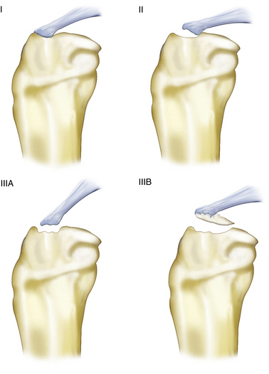
FIGURE 2-1 Meyers and McKeever classification of tibial intercondylar eminence fractures.
(Adapted from Lubowitz JH, Elson WS, Guttman D. Part II: Arthroscopic treatment of tibial plateau fractures: intercondylar eminence avulsion fractures. Arthroscopy. 2005;21:86-92.)
Associated injuries with fractures of the tibial eminence are common. Meniscus injuries are the most common injuries seen; however, these fractures may be associated with chondral and ligamentous injuries as well.11,12 In an unpublished study, we found an interposed intermeniscal ligament in 80% of types II and III injuries. This has profound implications for treatment strategies. In addition, tibial eminence fractures are also seen with tibial plateau fractures, specifically Schatzker types V and VI fractures.13
TREATMENT
The goal for management of tibial eminence fractures should be no different than for any other intra-articular fracture. Anatomic reduction and rigid fixation that allow for early range of motion should be the treatment for these fractures. Debate has ensued over anatomic reduction versus overreduction. It has been proposed that overreduction may result in excessive tension of the ACL, which results in limited knee range of motion.14 Others have countered this by stating that plastic deformation of the ACL occurs prior to the avulsion fracture and thus overreduction would result in a better outcome.9 Numerous studies have documented residual laxity in well-reduced tibial eminence fractures, and most have concluded that the laxity is not symptomatic.15–17 More studies are needed to answer the question of anatomic versus overreduction, but there is consensus that any displacement requires at least an anatomic reduction.
Management has been based on the Meyers and McKeever classification, with recommendations for immobilization in extension for type I fractures. Some controversy exists in regard to what degree the knee is to be extended for nonoperative management. Meyers and McKeever have recommended immobilization in 20 degrees of flexion.2,8 Similarly, Beaty and Kumar have recommended immobilization in 10 to 15 degrees of flexion.18 Fyfe and Jackson based their recommendations of flexing the knee to 30 to 40 degrees because the ACL is taut in extension and, with some flexion, the tension on the avulsion fragment would be less.19 These authors favor immobilization in full extension to avoid a flexion contracture, which can occur if the knee is kept in a flexed position. We encourage straight leg raises and quadriceps isometrics and allow full weight bearing, as tolerated, in a brace locked in full extension. The knee should not be immobilized in hyperextension because extensive stretch on the popliteal artery may result in a compartment syndrome.13 Regardless of the position of immobilization, close follow-up with radiographs weekly for 4 weeks should help confirm maintenance of reduction.
Treatment of type II fractures has been controversial. Closed reduction may be attempted by aspiration of the hemarthrosis and knee extension performed to allow the femoral condyles to help reduce the fracture.20

Stay updated, free articles. Join our Telegram channel

Full access? Get Clinical Tree


