Arthroscopic Rotator Interval Capsule Closure

Chapter 6


Arthroscopic Rotator Interval Capsule Closure*


Important Points


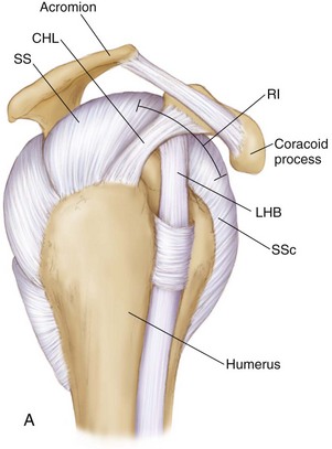
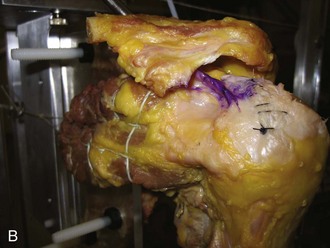
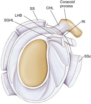
• The components of the RI include the coracohumeral ligament (CHL), the superior glenohumeral ligament (SGHL), the long head of the biceps (LHB) tendon, and a thin layer of joint capsule.


• The RI is a triangular space between the supraspinatus (SS) and subscapularis (SSc).


• The RI shape changes with internal and external rotation of the glenohumeral (GH) joint.


• A competent RI contributes to inferior shoulder stability via the CHL and an intact shoulder capsule (maintains negative intraarticular pressure).


• A sulcus sign that persists in external rotation (ER) is an indicator of RI insufficiency (of the CHL).


• Hyper-ER of the arm at the side (more than 90 degrees) also suggests incompetent anterior stabilization structures (possibly the RI).


• An open RI closure imbricates the CHL better than an arthroscopic closure; thus an open RI closure does not perform the same biomechanically as an arthroscopic RI closure, as both techniques generally repair different tissues in a different vector of closure.


• Volumetric reduction of the GH joint capsule may be achieved with adequate RI closure.


Clinical and Surgical Pearls


The rotator interval (RI) is a triangular space of the anterosuperior shoulder between the supraspinatus (SS) and subscapularis (SSc) tendons, containing both the coracohumeral ligament (CHL) and the superior glenohumeral ligament (SGHL) (Figs. 6-1 and 6-2). Although injuries to the RI capsule have been associated with increased glenohumeral translation and subsequent instability,1,2 its contribution to overall shoulder stability remains under debate. Several reports have suggested that RI capsular structures contribute to stability by resisting inferior and posterior glenohumeral translation36 and/or maintaining negative intraarticular pressure,7 whereas others have shown that surgical imbrication of the RI augments surgical correction of multidirectional and posterior instability.3,4,6,812 Previously, RI closure was commonly performed via open surgical techniques; however, recent all-arthroscopic techniques for RI closure have been described.13,14


The debate regarding RI closure is centered around the “circle concept” of the shoulder,15 which states that if the humerus is posteriorly subluxed, there must be an opposite and obligate injury to the anterior superior structures of the glenohumeral joint (the RI). However, several studies have refuted the circle concept theory,16 indicating no injury to the RI after posterior dislocation.


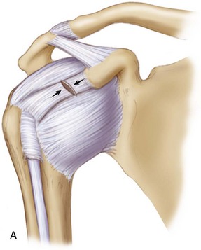
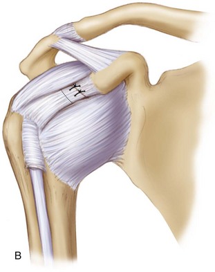
In addition, the premise of an open RI closure is not the same concept as an arthroscopic RI closure.3,14,17 As described by Harryman,3 open RI closure consistently imbricates the CHL from medial to lateral, which adequately restores inferior and posterior stability of the shoulder; however, this occurs at the expense of significant (30- to 40-degree) losses of external rotation (ER) at the side (Fig. 6-3). All-arthroscopic techniques have evolved to address the RI; however, the arthroscopic closure is fundamentally different from the open closure in direction of closure (arthroscopic: superior to inferior; open: medial to lateral and/or superior to inferior), in addition to differences in tissue imbricated (arthroscopic: RI capsule, SGHL to middle glenohumeral ligament [MGHL]; open: CHL or SS to SSc). Based on biomechanical evidence,12,14,1719 there are certain indications for an arthroscopic RI closure, including certain cases of anterior instability (in the setting of hyperlaxity) and revision anterior instability (to increase the bumper effect anteriorly), multidirectional instability with laxity and sulcus sign, and possible posterior or anterior instability in the setting of hyperlaxity. The purposes of this chapter are to review the operative indications and surgical technique for arthroscopic RI capsule closure in the setting of glenohumeral instability.


image


image


Figure 6-3 The coracohumeral ligament (CHL) is sectioned (A) and then imbricated by 1 cm (B), as was demonstrated by Harryman3 in an open rotator interval closure that shortened the CHL to improve inferior and posterior stability of the shoulder. This technique resulted in significant (30- to 45-degree) losses of external rotation at the side.


Preoperative Considerations


History


A thorough history is necessary in the evaluation of every patient with suspected RI pathology. RI pathology is most often seen with concomitant instability conditions of the shoulder, including anterior, posterior, and multidirectional instability. Therefore questions regarding baseline shoulder stability, including any history of traumatic instability events and/or chronic subluxation or dislocation events, should be asked. Information about any previous shoulder operations, especially stabilization procedures, should be noted. Given the complex anatomy of the RI, several concomitant structures may also be injured with an RI capsular lesion, and patients may report symptoms related to the labrum, CHL, biceps tendon, and rotator cuff. The diagnosis of any pathology in addition to lesions of the RI is thus crucial for preoperative planning and appropriate surgical management. The RI may be suspected to be involved in patients who have a history of ligamentous laxity, multiple recurrences of instability, and history of multidirectional instability or either anterior or posterior instability in the setting of hyperlaxity. Specifically, during the initial clinic visit, the clinician should inquire about the following:


Signs and Symptoms


Patients with RI capsular lesions often report diffuse shoulder pain, night pain, and the sensation of instability. RI pathology is often associated with the finding of shoulder instability, especially in the setting of hyperlaxity. Sensation of subluxation and/or dislocation, especially in the provocative position of abduction (ABD), and ER may be present. Hallmark symptoms that may be present include pain while carrying objects at the side (gallon of milk), paresthesias, and other findings suggestive of a sulcus sign or inferior laxity of the glenohumeral joint. Swelling is not commonly seen with these injuries and if present warrants a workup for other possible injuries, including articular cartilage defects. Similarly, strength and sensation are typically normal even in patients with significant RI capsule lesions, though limitations resulting from pain may be present.


Physical Examination


Isolated pathology of the RI is difficult to assess on examination, as findings are often vague and representative of anterior, posterior, and/or multidirectional instability. As in any shoulder examination, the appearance, strength, sensation, and range of motion (ROM) of the injured shoulder should be compared with those of the opposite shoulder in every patient with suspected RI capsule pathology. Particular attention should be paid to special tests for shoulder stability, because, as previously mentioned, RI lesions usually occur in the setting of shoulder instability. Physical examination findings suggestive of RI capsule lesions include the following:


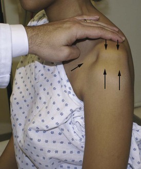
• Sulcus sign—downward traction of the arm causes inferior subluxation of the humeral head that does not resolve with ER of the shoulder (Fig. 6-4).


Stay updated, free articles. Join our Telegram channel

Sep 11, 2016 | Posted by in SPORT MEDICINE | Comments Off on Arthroscopic Rotator Interval Capsule Closure
Full access? Get Clinical Tree

Get Clinical Tree app for offline access