Arthroscopic Osteochondral Transplantation

CHAPTER 15 Arthroscopic Osteochondral Transplantation


Successful treatment of articular cartilage lesions is a complex clinical issue. The mechanical demands placed on articular cartilage in the knee °require it to withstand shear and compressive loads. Additionally, the patients themselves are often demanding, wishing to continue an already active lifestyle. This is further complicated by the limited healing potential of articular cartilage. The clinical outcome of a focal cartilage defect is highly dependent on the size, depth, pattern, and location of the injury. Multiple grading schemes have been described in an effort to quantify this, and hopefully will steer effective clinical treatment. However, no general consensus exists regarding the treatment of these lesions, and controversy continues to surround this topic.


Techniques for marrow stimulation, such as abrasion arthroplasty, drilling, and microfracture, produce fibrocartilage rather than native hyaline cartilage. Good short- and midterm results have been reported by various authors. However, inconsistent long-term results have perpetuated the interest in finding alternative methods for treating full-thickness chondral lesions. These include autologous chondrocyte implantation, autologous osteochondral transplantation, fresh osteochondral allografting, and bulk allografting, as well as a whole host of synthetics, scaffolds and first-, second-, and third-generation cell-based technologies.14 This chapter will focus on the COR system (DePuy Mitek; Raynham, Mass) for arthroscopic osteochondral transplantation.


The idea for the COR technique has its origins in an observation from anterior cruciate ligament (ACL) reconstruction that routine notchplasty is not associated with postoperative clinical sequelae. Because of this, the “notched” area (marginal cartilage in the lateral trochlear groove) of the knee can be used elsewhere, with minimal or no donor site morbidity. The design rationale for this technique is simple:


ANATOMY


Hyaline cartilage is has no nerve fibers and is avascular, deriving nutrition from its surrounding synovial fluid. Additionally, hyaline cartilage is composed mostly of matrix, with a relative paucity of live, active cellular material. Because of this, full-thickness articular cartilage defects have limited healing potential. When native healing of such defects occurs, fibrocartilage is formed in the defect. This is the goal of marrow-stimulating techniques, in which osseous bleeding at the defect initiates a cascade of fibrous scar healing. However, fibrocartilage is not as mechanically sound as hyaline cartilage under compressive or shear loads.


In adults, hyaline cartilage normally has a thickness of roughly 8 to 9 mm. As a person ages, the articular cartilage thins. The presence of pathology can accelerate this eroding process. To produce symptoms, articular cartilage must be thinned by more than 75% before subchondral nerve fibers can detect the change in contact surface pressure. However, overall size of the lesion also plays a role in this. A small chondral defect will not produce symptoms because the stress is distributed to the surrounding joint surface. Only when the cartilage defect becomes large enough, and thin enough will the patient become symptomatic.


To illustrate this point, Jason and Koh5 have examined the contact pressures across an articular surface with respect to a focal defect and a grafted defect. Various graft height mismatches were modeled (Fig. 15-1), with the results shown in Table 15-1. Not surprisingly, plugs that were flush to their articular surfaces most closely resembled the contact pressure of the normal intact articular surface. However, plugs countersunk by 0.5 to 1.0 mm also closely mimicked the native pressures. Thus, filling a defect to a near-congruent articular surface can reproduce native articular-surfaced pressures. This is the foundation of autogenous osteochondral transplantation.


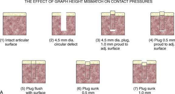
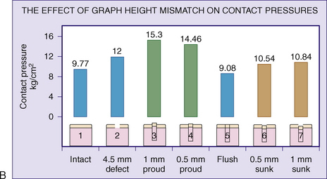
TABLE 15-1 Effect of Graft Height Mismatch on Contact Pressures


Condition of Intra-articular Joint Surface Contact Surface Pressure (kg/cm2)
Normal intact surface 9.77
Open 4.5-mm hole 12.00
Plug flush to the surface 9.08
Plug countersunk 0.5 mm to surface 10.54
Plug countersunk 1.0 mm to surface 10.84
Plug 0.5 mm proud to the surface 14.46
Plug 1.0 mm proud to the surface 15.30

Autogenous osteochondral transplantation, or mosaicplasty, is a technique in which multiple smaller grafts are harvested from a less weight-bearing portion of the knee and transplanted to the clinically relevant defect. The use of several small grafts has several advantages: (1) it allows the surgeon to reconstruct the natural anatomic contour lost by the defect; (2) it provides some hyaline surface structure to the reconstruction; (3) it limits fibrous cartilage growth to the gaps between the grafts and defect border; (4) it reduces donor site morbidity; (5) it is relatively inexpensive; and (6) the entire reconstruction can be done in a single surgery.


Because small chondral defects will not produce symptoms, the goal of osteochondral transplantation is to decrease the size of large defects functionally to become less clinically significant.


TREATMENT


Treatment should be tailored to various presenting factors. These include location and size of the lesion, age and weight of the patient, occupation, and type of sports involvement.


Indications and Contraindications


The COR system permits the arthroscopic harvesting of precisely sized articular cartilage–cancellous bone autografts from a suitable donor site, followed by the transplantation of these autografts to a precisely drilled defect site. The system can be used for open procedures if access to the defect or donor site is difficult. Indications for the procedure are single, full-thickness lesions at least 10 mm in diameter but not more than 35 mm in length or width. The depth of subchondral bone loss should not exceed 6 mm. Similar systems, although with some differences in design, are offered by the OATS system (Arthrex; Naples, Fla) and mosaicplasty (Smith and Nephew Endoscopy; Andover, Mass).


Indications for this procedure are as follows:


Contraindications include a history of degenerative joint disease or joint infection, intra-articular fracture, a diseased donor site, and multicompartment involvement. Additional physical contraindications relate to poor supporting bone at the recipient site, including a very large defect (more than 3 cm diameter), a deep defect (more than 6 mm deep), and extremely osteopenic subchondral bone stock. ACL disruption is not a contraindication, but concurrent ACL reconstruction is recommended in that case. Meniscal tears or prior surgeries on the lesion are not contraindications. No other area of significant chondral fibrillation or damage should be present. This technique is best performed on the femoral condyles and not on the tibial plateau.


Jun 19, 2016 | Posted by in MUSCULOSKELETAL MEDICINE | Comments Off on Arthroscopic Osteochondral Transplantation

Full access? Get Clinical Tree

Get Clinical Tree app for offline access