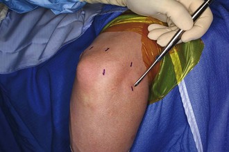
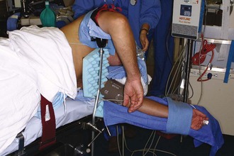
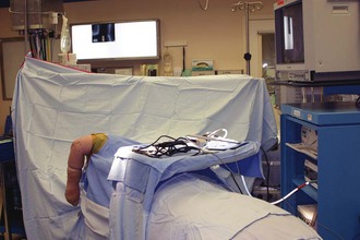
Chapter 44 Arthroscopy of the elbow is indicated for diagnostic therapeutic purposes in the setting of elbow arthritis and may be useful to treat many of the pathologic changes encountered with osteoarthritis, inflammatory arthritis, and posttraumatic conditions.1,2 Posttraumatic arthritis, osteoarthritis, hemophilic arthritis, and inflammatory arthritis may affect the elbow joint. Pain is a common complaint; however, patients may also report stiffness, weakness, instability, mechanical symptoms, or cosmetic deformity.3 A description of the degree and direction of motion loss and presence and occurrence of pain is an important part of the history. A history of trauma may be elicited from patients. The most common injury that may cause posttraumatic arthritis is a comminuted intra-articular fracture resulting in articular incongruity.3 Posttraumatic contractures caused solely by capsular fibrosis may be secondary to hemarthrosis from various traumatic causes. The patient should be asked whether he or she had physical therapy, whether it was painful or relatively benign, and whether splints had been used for short- or long-term sessions. Osteoarthritis involving the elbow is most commonly seen in the dominant arm of men with a history of heavy labor, weightlifters, and throwing athletes.3 Crutch ambulators may also be more prone to this condition. These patients are commonly in the third to eighth decades. Patients typically report pain at extremes of motion with loss of terminal extension more than loss of flexion. Patients with osteoarthritis who may benefit from arthroscopic debridement typically lack significant pain in the mid-arc of motion; rather, pain will be noted more at the end arc of flexion and/or extension owing to impinging terminal osteophytes. The presence of mid-arc of motion discomfort or pain throughout the arc of motion may be indicative of more widespread joint changes that may not respond as well to arthroscopic debridement. Symptomatic rheumatoid arthritis of the elbow fortunately is becoming less common with the advent of disease-modifying agents but can be quite debilitating when it does occur. Both elbows are commonly involved, and patients initially have pain and swelling from synovitis and effusion. More severe involvement with loss of bone and failure of the soft tissues around the elbow joint may cause instability, which may accelerate articular destruction as a result of subluxation or malalignment.4,5 Patients with hemophilia commonly develop arthropathy of the elbow. This appears to be related to some threshold of clinical or subclinical intra-articular bleeding, which seems to result in potentiation of an inflammatory cascade leading to worsening synovial proliferation and hemarthroses, with subsequent arthritis. Patients may report recurrent intra-articular bleeds caused by proliferative synovitis, or the secondary arthritic changes that ensue.6 Placement of the patient in the lateral decubitus position allows excellent access to the elbow joint (Fig. 44-1). The arm is placed in a padded arm holder that is attached to the side of the table. A low-profile elbow arm holder specifically designed for this purpose may be used. A nonsterile tourniquet is then placed on the arm at the level of the arm holder, and the arm is firmly secured to the arm holder. This facilitates arthroscopy by keeping the arm stable, just as a knee holder maintains stability during knee arthroscopy. The elbow should be positioned slightly higher than the shoulder. This allows 360-degree exposure of the elbow joint, eliminating the potential for impingement of the arthroscope or other instruments against the side of the body (Fig. 44-2). All portal sites are marked before surgery or insufflation of the joint, when the elbow is not distended or edematous and palpation of osseous landmarks is more precise (Fig. 44-3). Surface landmarks that should be marked include the lateral epicondyle, medial epicondyle, radial head, capitellum, ulnar nerve, and olecranon. The ulnar nerve is palpated to determine its location and to ensure that it does not subluxate from the cubital tunnel. Figure 44-3 Intraoperative view of portal placement. The retractor is demonstrating the standard anterolateral portal.
Arthroscopic Management of the Arthritic Elbow
Preoperative Considerations
Surgical Technique
Surgical Landmarks, Incisions, and Portals

![]()
Stay updated, free articles. Join our Telegram channel

Full access? Get Clinical Tree


Arthroscopic Management of the Arthritic Elbow


