Arthroscopic Management of Elbow Stiffness

Chapter 42


Arthroscopic Management of Elbow Stiffness


Clinical and Surgical Pearls


Loss of mobility represents the most common complication of elbow trauma. The predisposition of the elbow to develop posttraumatic contracture has been attributed to several factors, including the intrinsic congruity of the ulnohumeral articulation, the presence of three articulations within a synovium-lined cavity, and the intimate relationship of the capsule to the intracapsular ligaments and extracapsular muscles.13


Several authors have evaluated the elbow motion necessary to complete daily activities; these studies have reported that a majority of activities could be reasonably performed within a functional arc of 100 degrees (30 to 130 degrees) of flexion and extension of the elbow and 100 degrees of rotation of the forearm (50 degrees each for pronation and supination).4 The inability of the elbow to achieve this motion after the occurrence of trauma may result in substantial impairment of upper extremity function, especially with the loss of elbow flexion and forearm supination, which are difficult to accommodate for.


Nonoperative management remains the initial modality of treatment and typically includes static splinting as well as structured rehabilitation and physical therapy dedicated toward regaining functional range of motion. These modalities are particularly useful for the first 6 to 12 months postinjury but become less successful in cases of more remote trauma. For patients whose elbow contracture is refractory to conservative measures, surgical debridement and release of the elbow are offered in an effort to restore functional motion of the joint. Although open approaches have been classically described for the surgical treatment of the posttraumatic elbow contracture, arthroscopic techniques have recently emerged as a less invasive alternative for the treatment of elbow stiffness and demonstrate similar efficacy within the literature.


Preoperative Considerations


History


It is imperative for the practitioner to determine the extent to which the loss of elbow motion compromises a patient’s functional capabilities. The magnitude of functional impairment, rather than absolute loss of motion, ultimately directs management decisions when treating the patient with posttraumatic contracture of the elbow.


The details surrounding the initial injury as well as mechanism of trauma are important aspects of the history. Many of these patients will have undergone previous surgical treatment, and it is critical to obtain and review previous operative documentation, especially when arthroscopic treatment is being considered. Complications related to initial treatments, including infection, neurologic deficits, or other ipsilateral limb injuries, all potentially influence future management. In addition, details regarding the patient’s progress or, more important, lack of progress with structured rehabilitation should be elicited during the history taking.


Physical Examination


Physical examination begins with inspection of the entire upper extremity, specifically evaluating for deformity, swelling, and muscle atrophy while noting the location of any previous surgical incisions that would influence further surgical planning. Range-of-motion evaluation should include the hand, wrist, forearm, and elbow and should be compared with the contralateral, unaffected extremity. In the posttraumatic setting, loss of extension is more common than loss of flexion.


A careful neurovascular examination is essential, especially during the evaluation of ulnar nerve function. Residing in the cubital tunnel and adjacent to the medial joint capsule, the ulnar nerve may become entrapped in scar tissue along the medial elbow after trauma, which may result in posttraumatic ulnar neuropathy. Traction ulnar neuritis of the elbow may manifest as medial elbow pain, and patients may report sensory changes in an ulnar nerve distribution, particularly with elbow flexion. Patients with posttraumatic ulnar neuropathy may simply have loss of flexion and medial elbow pain in the absence of overt symptoms of ulnar neuropathy; thus a meticulous neurovascular evaluation, including assessment of two-point discrimination, pinch strength, and intrinsic muscle function, are essential to document the preoperative function of the ulnar nerve. Elbow stability must also be assessed, in particular to rule out subtle posterolateral rotatory instability as a cause of the patient’s complaints of stiffness.


Imaging


Standard plain radiographs of the elbow are typically obtained and include anteroposterior, lateral, and oblique projections. Stress radiographs should be considered if elbow instability is suspected. Computed tomography (CT) is frequently performed, especially when the presence of heterotopic ossification (HO) or intra-articular loose bodies is suspected or if bony deformity or malunion is suspected in the setting of previous fracture. Three-dimensional CT reconstruction is typically helpful in further delineating bony and articular anatomy. Advanced imaging is important in documenting ulnohumeral joint congruency as well as any osseous impingement secondary to overgrowth in the olecranon or coronoid fossae that would directly limit motion. We have found magnetic resonance imaging (MRI) to have a limited role.


Indications and Contraindications


Nonoperative management is routinely initiated after the initial evaluation of the patient with a posttraumatic elbow contracture and typically includes a course of structured rehabilitation and the use of preadjusted static braces for passive progressive stretch of the soft tissues. Supervised therapy helps ensure compliance and can also be used to document progress with therapy.


Surgical management is indicated for patients who continue to experience significant loss of mobility with resultant impairment of upper extremity function and limitation with daily activities. Although a flexion contracture of at least 25 to 30 degrees and/or less than 110 to 115 degrees of active flexion was historically reported as an indication for surgical contracture release, surgical management may still be offered to certain patients who may require full or nearly full motion for specific lifestyle and employment demands. Patients typically must be at least 3 to 4 months removed from injury to allow them to achieve “tissue equilibrium,” with maximal resolution of posttraumatic swelling and inflammation. Most important, patients must be willing to comply with the required extensive program of postoperative therapy, as operative outcomes depend on diligent participation in a structured rehabilitation program. This is especially true for adolescents, who may not be dedicated to improving their elbow motion.


Relative contraindications to arthroscopic elbow release include severe elbow contractures with minimal joint motion, prior ulnar nerve transposition surgery, and the presence of significant heterotopic bone; these patients are more reliably treated with extensive open debridement of the elbow to restore motion and protect the ulnar nerve. If there has been previous medial elbow surgery or ulnar nerve transposition and one is considering arthroscopic release, the ulnar nerve should be isolated and protected through a medial exposure before arthroscopy. If the ulnohumeral joint shows marked degenerative changes, a simple release of the joint may not lead to improved motion and may exacerbate pain in an arthritic joint. If advanced posttraumatic arthritis is observed in the ulnohumeral articulation, salvage-type procedures are often required if surgery is undertaken.5


Subtle elbow instability commonly manifests as loss of motion after elbow fracture or dislocation; accordingly, special attention should be devoted toward evaluating elbow stability either with stability testing on physical examination or with stress radiographs. If instability is present, then treatment typically would include ligament reconstruction with or without capsular release. The stiff and unstable elbow is a particularly challenging condition to treat. The priority is to achieve stability and to later restore motion, typically in a staged procedure.


Surgical Technique


Arthroscopic release of a posttraumatic elbow is a technically demanding procedure that requires intimate knowledge of intracapsular elbow anatomy as well as advanced skills in elbow arthroscopy. Multiple portals are required, and diligent fluid management is essential, especially after capsulectomy when joint distention is more difficult. The use of joint retractors is typically required to achieve adequate visualization and facilitate appropriate surgical debridement.610


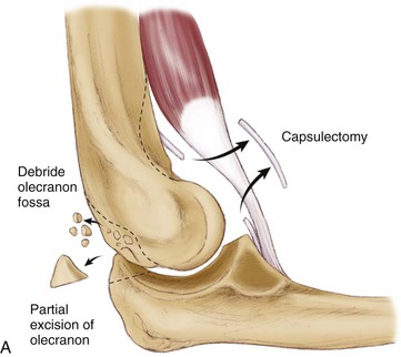
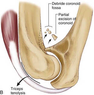
From a purely mechanical standpoint, to improve elbow extension, posterior impingement must be removed between the olecranon tip and the olecranon fossa. Anteriorly, tethering soft tissues such as the anterior joint capsule and any adhesions between the brachialis and the humerus must be released. Similarly, to improve elbow flexion, the surgeon must release any soft tissue structures posteriorly that may be tethering the joint. They include the posterior joint capsule and the triceps muscle, which can become adherent to the humerus. The surgeon must remove any bony or soft tissue impingement anteriorly, including any soft tissue overgrowth in the coronoid and radial fossae. There must be a concavity above the humeral trochlea and capitellum to accept the coronoid centrally and the radial head laterally for full flexion to occur (Fig. 42-1).


Anesthesia and Positioning


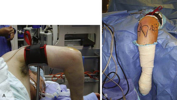
We typically favor a regional block, although general anesthesia may also be considered. We then position the patient in the lateral decubitus or prone position with the affected extremity facing up and all bony prominences well padded. A rolled blanket or elbow stirrup attachment for the operative table is positioned underneath the arm, allowing the elbow to move from 90 degrees of flexion to full extension. The video monitor, shaver system, camera control unit, and light source are positioned such that the surgeon has a clear view of the monitor. After the extremity has been sterilely prepared and draped up to the axilla, the hand and forearm are wrapped with an elastic bandage to limit fluid extravasation and a sterile pneumatic tourniquet is placed as proximally as possible around the arm (Fig. 42-2).


Stay updated, free articles. Join our Telegram channel

Sep 11, 2016 | Posted by in SPORT MEDICINE | Comments Off on Arthroscopic Management of Elbow Stiffness

Full access? Get Clinical Tree

Get Clinical Tree app for offline access