Arthroscopic Management of Elbow Osteochondritis Dissecans Lesions


Arthroscopic Management of Elbow Osteochondritis Dissecans Lesions


Guillem Gonzalez-Lomas

Neal S. ElAttrache


INTRODUCTION

The incidence of sport-specific injuries in young athletes has increased with earlier and more rigorous young athlete participation in sports. The radiocapitellar compartment of the young athlete’s elbow withstands significant stresses during repetitive activities (such as throwing) or during sports that convert the elbow joint into a weight-bearing joint (such as gymnastics) (1). Specifically, lateral compartment compression can lead to Panner diseases (osteochondrosis) in the very young, preadolescent (6-10-year-old) patient or capitellar osteochondritis dissecans (OCD) in the adolescent or young adult (2, 3, 4, 5). OCD may in turn generate loose bodies. This chapter describes Panner disease and OCD, and describes a treatment algorithm, including detailing their arthroscopic management.


PANNER DISEASE

In 1927, Hans Jessen Panner described “osteochondrosis” of the capitellum, remarking on its similarities to Legg-Calve-Perthes of the hip (6). Like other ostochondroses, it consists of noninflammatory disordered endochondral ossification. Its specific etiology and relationship to OCD remain debatable. It is generally accepted, however, that abnormal radiocapitellar compressive forces during a period of vulnerability predispose children to it (2,5,7). Etiologically, it may result from the combination of an avascular insult (likely related to the capitellum’s predominantly end-artery supply) and repetitive microtrauma (8).


Epidemiology

Panner disease predominantly affects boys younger than age 10 (9). Young boys tend to be predisposed to it for two reasons. One, compared to girls, they have a delayed appearance and maturation of their secondary growth centers. Two, boys traditionally are more prone to trauma during the more aggressive early childhood activities they select (7). This may change as more girls become involved in higher risk athletic activities at younger ages. Although Panner’s can be confused with OCD, and the age of onset may overlap, it distinguishes itself by three epidemiological characteristics. One, Panner disease does not share the strict association with repetitive throwing that OCD does; Two, it is usually self-limiting. And three, it resolves without any long-term sequelae.


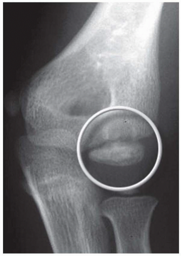
FIGURE 4.1 Panner disease. AP radiograph of the left elbow demonstrating fragmentation and lucency of the capitellum (circle) near the chondral surface.


Presentation

Patients with Panner disease will initially present complaining of pain and stiffness in the elbow, relieved by rest. On physical exam, they will have poorly localized tenderness over the lateral elbow. Radiographs will initially show fissuring, lucencies, fragmentation, and irregularity of the capitellum (shown in Fig. 4.1), particularly near or at the chondral surface. Subsequent films, taken at 3 to 5 months, will demonstrate larger radiolucent areas followed by reossification of the bony epiphysis with a corresponding resolution of symptoms. In 1 to 2 years, the epiphysis regains its contour, usually without flattening (4). It should be noted that, as in Legg-Calve-Perthes, radiographs often lag behind clinical symptoms. MRI can also document the extent of the lesion. Typically, edema is localized to the chondral surface with less involvement of the subchondral bone, as compared to OCD (Fig. 4.2).


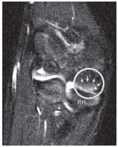
FIGURE 4.2 Panner disease, as seen on T2-weighted MRI. Circle surrounds Panner lesion, demarcated by small arrows, opposite radial head (RH). Notice the more typical finding of edema adjacent to the capitellar chondral surface, rather than deeper in the subchondral bone as in OCD.


Treatment

Treatment involves complete rest from the activity in question and administering modalities such as ice and anti-inflammatory medication. The elbow may occasionally need to be immobilized for 3 to 4 weeks to control symptoms. Symptoms usually resolve within 6 to 8 weeks, although they occasionally persist for months, and activities should be reinstituted progressively and as tolerated. The condition has excellent long-term prognosis, although in some patients there may be a slight residual flexion contracture.


OSTEOCHONDRITIS DISSECANS

OCD of the capitellum is a noninflammatory degeneration of subchondral bone occurring in the context of repetitive trauma to the lateral compartment of the elbow. Panner disease and OCD may represent two different stages of the same disorder (4). The two conditions, however, do differ in certain characteristics: age of onset, etiology, and natural history. One, while Panner disease affects children under 10, OCD victimizes older athletes between ages 11 and 15 (10). Two, unlike Panner disease, OCD is thought to be directly linked to repetitive trauma. Three, OCD is not always self-limiting. If left unaddressed, it results in profound destruction of the capitellum (10).


Etiology

OCD arises from repetitive and excessive compressive forces generated by either large valgus stresses on the elbow during throwing or racket swinging or from constant axial compressive loads on the elbow such as those endured by gymnasts (10, 11, 12). Specific risk factors predispose patients to the condition. In the case of baseball players, throwing sliders and breaking pitches, throwing more than 600 pitches per season, and increased age of the athlete increase the risk of developing OCD (12). In female gymnasts, overtraining involving excessive handstand maneuvers has been linked to OCD (13,14). Other risk factors include genetic predisposition and the tenuous end-artery vascular supply to the capitellum. In the young adult population, the capitellum is supplied by two end arteries coursing from posterior to anterior which are branches of the radial recurrent and interosseous recurrent arteries (Fig. 4.3) (15). As a result of the longitudinal blood supply to the capitellar epiphyseal plate and minimal collateral circulation in the area, blood flow to the capitellum may be disrupted by both repetitive microtrauma resulting in an avascular state, and a single traumatic event leading to posttraumatic subchondral bone bruises (16,17).


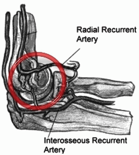
FIGURE 4.3 Capitellar blood supply. In the young adult population (under age 20), the radial recurrent and interosseous recurrent arteries give off branches that course from posterior to anterior and supply the capitellum (inside red circle). This end-artery blood supply makes the capitellum susceptible to an avascular insult.


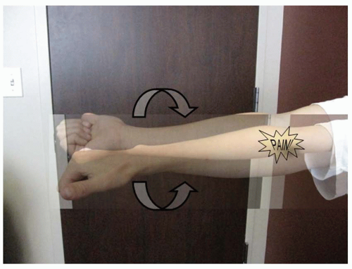
FIGURE 4.4 Radiocapitellar compression test. Pain in lateral elbow when the extended arm is pronated and supinated.


Presentation

Patients with OCD will initially present complaining of pain and stiffness in the elbow, relieved by rest. If left unaddressed, the symptoms may progress to “locking” or “catching” due to intraarticular loose bodies. Physical examination tends to be remarkable for poorly localized lateral elbow tenderness over the radiocapitellar joint. Loss of range of motion with a 15 to 20 degrees flexion contracture is common. Loss of extension is more common than loss of flexion. Radiocapitellar joint provocative maneuvers include the active radiocapitellar compression test (Fig. 4.4). A positive test elicits pain in the lateral compartment of the elbow when the patient pronates and supinates the forearm with the arm in extension.


Imaging

Anterior-posterior in full extension, anterior-posterior in 45 degrees of flexion, and lateral views of the elbow should be obtained. Radiographs may be negative early in the disease process. As the condition progresses, flattening and sclerosis of the capitellum, typically on its anterolateral aspect, will become apparent. Irregular areas of lucency and intraarticular loose bodies also appear. Both the capitellar lesions of OCD and medial-sided epicondylar fragmentation are best seen on an AP at 45 degrees elbow flexion (Fig. 4.5). In suspected OCD, an MRI should always be obtained. It will detect bone edema early in the disease process (18). An MRI arthrogram can further delineate the extent of the injury. The contrast can show separation of a detached or
partially detached piece from subchondral bone (Fig. 4.6). Peiss et al. (19). felt that fragment enhancement (seen in Fig. 4.7B) (as opposed to the perifragment enhancement seen in Fig. 4.6) denotes viability and may be a reasonable indication for nonoperative treatment. They also suggested that enhancement of the fragment-subchondral bone interface is caused by vascular granulation tissue, indicating instability and requiring operative intervention. Of note, it is critically important to distinguish “pseudolesions,” appearing on the posteroinferior
junction of the articular and nonarticular portions of the capitellum, from OCD which almost always presents on the anterolateral aspect. In addition, whether or not the capitellar physis is open or closed should be noted.


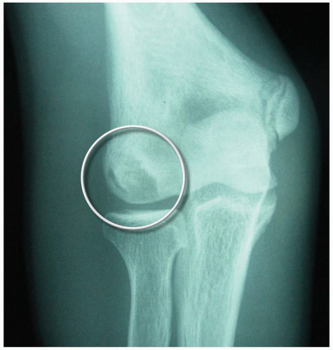
FIGURE 4.5 AP radiograph at 45 degrees flexion. OCD lesion (circle) is seen more clearly with elbow flexed to 45 degrees.


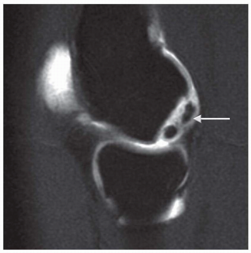
FIGURE 4.6 MR arthrogram showing contrast surrounding unstable OCD fragment (arrow).


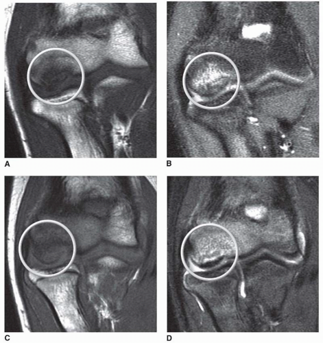
FIGURE 4.7 Stage 1 OCD lesion progress. Stable, intact, nondisplaced fragment (circles) with abnormal signal on coronal slices in (A) T1 and (B) T2 sequences. After 6 months of conservative management (C) T1 and (D) T2 sequences show reconstitution of subchondral bone in area the lesion. The patient was symptom free at the 6-month follow-up.


TABLE 4.1 Classification and Treatment of Capitellar Osteochondritis Dissecans Lesions


Stability


Stage


MRI Findings


Arthroscopic Findings


Treatment


Stable


I


Normal XR
T1 abnormal
T2 normal


Intact articular cartilage
Subchondral bone edema but structurally sound


1.


Hinged elbow brace: 3-6 wk


2.


PT


3.


NSAIDs


4.


Follow-up XR and or MRI at 3-6 mo


Unstable


II


Abnormal XR
T1/2 abnormal
Contrast shows margin around lesion


Partially detached fragment
Cartilage fracture
Subchondral
bone collapse
Lateral buttress involved: poorer prognosis


1.


Acute: Consider fragment fixation, but higher success treating as chronic (below).


2.


Chronic:


a)


<6-7 mm lateral buttress involved/radial head does NOT engage: Fragment removal + microfracture/drilling


b)


>6-7 mm lateral buttress involved/head engages: Removal + osteochondral autograft/synthetic graft


III


Loose bodies


Completely detached
Loose bodies


1.


Loose body removal


2.


Treat as Stage II


Associated radial head OCD


Any of the above


1.


<30% radial head involvement: Treat as Stage II


2.


>30%: No osteochondral grafting; Microfracture drilling ok


Management

Management of OCD lesions is based primarily on the status and stability of the overlying cartilage. The size and location of the lesion and the patency of the capitellar growth plate also influence decision making (20, 21, 22). In order to guide treatment, detailed classification systems based on radiographic (23,24) and arthroscopic (20) findings have been delineated (25,26). We have chosen to simplify these algorithms into a succinct, three-stage classification that provides a template for management. Table 4.1 illustrates the classification.


Stage 1

In Stage 1, the osteochondral fragment is intact, stable, and nondisplaced. Radiographs are often negative. Signal on MRI is variable, typically abnormal on T1 and normal on T2, although T2 signal may also be abnormal. Figure 7A and B

Only gold members can continue reading. Log In or Register to continue

Stay updated, free articles. Join our Telegram channel

Jun 13, 2016 | Posted by in ORTHOPEDIC | Comments Off on Arthroscopic Management of Elbow Osteochondritis Dissecans Lesions

Full access? Get Clinical Tree

Get Clinical Tree app for offline access