CHAPTER 14 Arthroscopic Labral Repair
Introduction
Our understanding of hip labral pathology as a cause of hip pain is evolving, and the various treatment interventions available are also in flux. The exact prevalence of acetabular labral tears in the general population is unknown. However, when investigating the incidence of these injuries among athletes who present with groin pain, a clinical assessment of 18 patients who presented to a sports clinic with complaints of groin pain and an age range of 17 to 48 years revealed that, in 4 of the 18 athletes (22%), a labral tear was documented with the use of magnetic resonance arthrography. On the basis of this analysis, it appears that an acetabular labral tear is certainly not uncommon and that treatment algorithms need to be delineated.
The current indications for labral repairs are somewhat variable. Although the labral tear as a result of a single traumatic event is the clearest indication for repair, this is a rare entity in clinical practice. More often, the inciting event is not obvious and chronic repetitive injuries leading to attritional tears are at fault, whereas in other cases femoroacetabular impingement or other bony variants may be at fault. It is important to realize not only that the labral tear should be treated but also that the underlying problems need to be addressed, or else the long-term outcomes will be poor.
Basic science
The hip labrum is a fibrocartilaginous structure that surrounds the rim of the acetabulum in a nearly circumferential manner. It is contiguous with the transverse acetabular ligament across the acetabular notch inferomedially. The labrum is widest in the anterior half and thickest in the superior half, and it merges with the articular hyaline cartilage of the acetabulum through a transition zone of 1 mm to 2 mm. It is attached to the edge of the bony acetabulum via a thin tongue of bone that extends into the tissue via a zone of calcified cartilage, and it adheres directly to the outer surface of this bony extension without a zone of calcified cartilage. A group of three or four vessels are located in the substance of the labrum on the capsular side of this extension. The structure is separated from the hip capsule by a narrow synovial-lined recess, which is variable in size. In general, the vascular supply of the adult hip labrum is poor, and regional differences in vascularity exist. Kelly and colleagues used cadaveric injection studies to define the vascularity of this area, and they demonstrated that the capsular portion of the labrum is significantly more vascular than the articular side, with only the peripheral third of the labrum having nutrient vessels. Extrapolating from our understanding of the healing capacity of the meniscus, repair strategies should be considered for tears that involve only the peripheral labrum.
The labrum functions to enhance the stability of the hip by maintaining the negative intra-articular pressure within the joint as well as by increasing congruity. With the use of a poroelastic finite element model, it has been determined that the labrum functions to provide structural resistance to the lateral motion of the femoral head within the acetabulum. Furthermore, it has been demonstrated that the labrum also functions to decrease contact pressures within the hip and to decrease cartilage surface consolidation. This effect is a result of maintaining the articular fluid in contact with the weight-bearing cartilage via a joint-sealing effect.
Diagnosis of labral tears of the hip
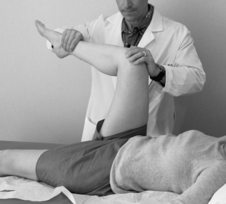
The clinical presentation of patients with a tear of the labrum is variable, and, as a result, the diagnosis is often missed initially. Burnett and colleagues reported about a series of 66 patients in whom the diagnosis of a labral tear had been made by arthroscopy. In this series, the mean time from the onset of symptoms to diagnosis was 21 months. An average of 3.3 health care providers had seen each patient before the diagnosis of a labral tear was made. Groin pain was the most common complaint (92%), with the onset of symptoms most often being insidious. A positive “impingement sign” occurred in 95% of patients in this series; this sign consists of groin pain with flexion, adduction, and internal rotation of the symptomatic hip (Figure 14-1). Therefore, in young, active patients who present with complaints of groin pain, with or without a history of trauma, the diagnosis of a labral tear of the hip should be suspected and investigated further.
Imaging and diagnostic studies
Standard radiographs (weight-bearing anteroposterior pelvis and frog-leg lateral) are critical as part of the initial assessment to evaluate the patient for arthritis and femoroacetabular impingement and also for more subtle dysplasias. Multiple studies have demonstrated the superior accuracy of magnetic resonance arthrography (MRA) as compared with standard magnetic resonance imaging for the diagnosis of labral tears. Intra-articular gadolinium has been shown to improve the sensitivity of diagnosing labral pathology from 25% to 92% with the use of a small field of view. Therefore, when clinical suspicion of a hip labral tear exists, MRA with a small field of view is the study of choice.
An intra-articular lidocaine injection is useful for situations in which the diagnosis of labral pathology is equivocal or in which a tear has been diagnosed by MRA but it is uncertain whether symptoms are related to this finding. Similar to its use for diagnosing external impingement of the shoulder, if patients experience relief from symptoms after the injection, the diagnosis of pain as a result of hip intra-articular pathology is more certain. Although this does not confirm that the labral tear is the problem, it certainly makes the case that intra-articular pathology is at least somewhat responsible for the symptomatology.
Surgical technique
The decision to proceed with operative intervention should be heavily weighed with regard to refractory mechanical symptoms. The majority of labral tears are treated with debridement; however, some tears are amenable to arthroscopic repair. Because the blood supply to the labrum enters from the adjacent joint capsule, peripheral tears have healing potential, and repairs should be considered if this pattern is encountered.
Technique for Arthroscopic Labral Debridement
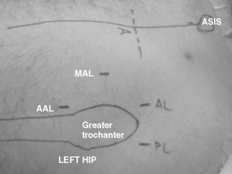
The procedure can be performed with the patient in either the supine or lateral position, depending on the level of comfort of the surgeon. The procedure should employ both a 30-degree and a 70-degree arthroscope for a thorough assessment of both the labrum and associated pathology. Modified arthroscopic flexible instruments, extended shavers, and hip-specific instrumentation should be available to improve access to all areas of the hip joint. In addition, the positioning of instruments should be done in the proper portals, with consideration for the anatomic structures near the hip joint (Figure 14-2).

Figure 14–2 The standard arthroscopic portals employed for access to the hip joint (in this case, the left hip) with the patient in the supine position. The accessory mid-anterolateral portal is useful as a result of its appropriate angle of approach to the acetabular bone. A, Anterior portal; AAL, accessory anterolateral portal; AL, anterolateral portal; ASIS, anterosuperior iliac spine; PL, posterolateral portal; MAL, mid-anterolateral portal.
A diagnostic arthroscopic examination of the central compartment can be performed systematically to evaluate not only the labrum from anterior to posterior but also to locate possible cartilage lesions on both the acetabular and femoral sides. In addition, the integrity of the ligamentum teres should be assessed; this area can be a source of pain as a result of impingement of the soft tissues between the femoral head and the acetabulum. Any loose bodies should be noted and their source identified. Finally, an assessment should be made of any obvious capsular redundancy or laxity.
Many patients will have a significant synovitis associated with the labral tearing, and an effort should be made to resect some of the inflamed tissue to improve visualization of the joint and to decrease the associated pain. This should be undertaken with a radiofrequency probe to decrease the potential for bleeding and the subsequent compromise of the surgical field.
The goal of the surgical procedure should be to preserve as much native tissue as is technically feasible while resecting the degenerative or damaged material. This is important to maintain the labrum’s role as a secondary joint stabilizer and to minimize the potential for arthrosis. Frayed tissue should be debrided with the use of either motorized shavers or radiofrequency probes. It is important to delineate the areas of abnormal tissue that are identified both on radiographs (in the form of perilabral calcifications) and with magnetic resonance imaging or MRA (abnormal signal intensity) to thoroughly address the labral pathology.
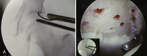
Adjacent cartilage damage should be searched for and thoroughly addressed. Superficial lesions can be gently debrided with mechanical shavers and perhaps stabilized with the use of radiofrequency probes. Grade IV Outerbridge lesions should be managed with a thorough debridement down to a bleeding bed and by preparation with microfracture awls (Figure 14-3).

Figure 14–3 Chondroplasty of an articular cartilage lesion. A, Chondroplasty awl used for the preparation of significant chondral injuries of the acetabular and femoral cartilage. B, Final area of preparation with an obvious bleeding bed.

Stay updated, free articles. Join our Telegram channel

Full access? Get Clinical Tree