Arthroscopic Capsular Plication and Thermal Capsulorrhaphy

CHAPTER 15 Arthroscopic Capsular Plication and Thermal Capsulorrhaphy


Introduction


The hip is an inherently stable articulation between the femoral head and the acetabulum. Unlike the shoulder, the primary component of hip stability is determined by its constrained osseous anatomy. Although traumatic hip subluxations and dislocation are well documented, recently there has been heightened interest in atraumatic instability as a source of recalcitrant hip pain and symptoms. In these cases, redundant or incompetent capsular–labral structures lead to microinstability. There is a dynamic and transient incongruency or subluxation within the femoroacetabular articulation that results in abnormal force distributions across the hip joint and ultimately in worsening capsular redundancy and injury, chondrolabral injuries, and femoral neck impingement at high flexion angles (i.e., secondary impingement). This ignites clinical symptoms and starts a cascade of pathologic events that result in hip pain, stiffness, flexion contractures, labral pathology, and degeneration of the hip. The causes of hip instability and capsular laxity are multifactorial and include such things as intrinsic ligamentous laxity, connective tissue diseases, overuse or repetitive activities, iatrogenic injuries, subtle hip subluxation injuries, and prior dislocations.


The mainstay of treatment for capsular laxity and atraumatic instability has been conservative. There have been a few reports of open capsular plication as a method of treatment, but these have mostly been for posttraumatic instability. Recently, improved understanding and imaging of the prearthritic hip has led to the development of arthroscopic treatment approaches. Early experience was primarily focused on arthroscopic thermal capsular shrinkage and capsulorrhaphy, which produced promising early results. However, because thermal capsulorrhaphy has recently fallen out of favor for addressing shoulder instability because it can result in collagen disruption, chondrolysis, and high long-term failure rates, there has been gradual movement toward arthroscopic hip capsular plication. This burgeoning technique provides the advantages of a reproducible and durable capsulorrhaphy with the desired amount of plication controlled by the surgeon without the potentially adverse affects of thermal shrinkage. We present here a review of atraumatic hip instability and capsular laxity, and we will discuss their relevant anatomy, evaluation, imaging studies, and management principles as well as arthroscopic techniques of capsular plications in both the central and peripheral compartments.


Basic science


The hip is a constrained diarthrodial joint. The femoral head is an approximation of a sphere, and, under normal osseous parameters, the acetabulum covers approximately 170 degrees of the femoral head. The labrum is a fibrocartilage structure that further deepens the acetabulum. It functions to enhance stability by establishing a negative intra-articular pressure within the hip joint, preserving joint congruity, and limiting the fluid expression that acts as an important sealing function. The labrum also plays a role in helping to contain the femoral head in extremes of range of motion, especially in flexion. Its role in providing rotational stability is still unknown.


The surrounding capsular envelope consists of three ligaments: the iliofemoral ligament (i.e., the Y ligament of Bigelow), the pubofemoral ligament, and the ischiofemoral ligament (Figure 15-1). The iliofemoral ligament has a medial and lateral limb proximally, and distally it forms a deep circular band that surrounds the femoral neck in a leash-like fashion; this area is called the zona orbicularis. This “Y ligament” is the strongest, and it prevents the anterior translation of the hip during extension and external rotation when its fibers tighten. In flexion, these fibers loosen, which leads to a “screw home” effect in full extension. The pubofemoral ligament is slightly inferior to the iliofemoral ligaments, and it also controls external rotation in extension. The ischiofemoral ligament is a posterior structure that controls internal rotation in flexion and extension. Other secondary hip stabilizers include the ligamentum teres and the psoas tendon, which may provide important stability in cases of dysplasia or static ligament deficiencies.


The causes of chronic atraumatic hip instability and capsular laxity are multifactorial. Primary causes include milder forms of intrinsic ligamentous laxity as well as more extreme cases of connective tissue diseases such as Marfan syndrome or Ehlers-Danlos syndrome, with which patients may be able to voluntarily or habitually dislocate their hips. The more common (but less well-recognized) secondary causes are overuse and repetitive activities such as golf, dancing, gymnastics, and martial arts. In these cases, there is repetitive hip rotation with an axial load. Other secondary causes include iatrogenic injuries and subtle forms of trauma, including hip subluxations and even prior dislocations.


The last type of atraumatic hip instability involves osseous anatomic abnormalities on either the acetabular side or the femoral side. Inclination, version, and other osseous parameters of the weight-bearing surface affect the soft-tissue structures that surround the hip. For cases in which there is a deficiency of the bony acetabulum (dysplasia) or excessive femoral anteversion or valgus, there is more reliance on the surrounding soft-tissue structures. McKibbin has quantified this with an index of combined femoral and acetabular version that predicts increased stress to the anterior capsulolabral structures. He defined the McKibbin index as the sum of the angles of femoral and acetabular anteversion, with a total of more than 60 denoting severe instability.


Brief history and physical examination


The diagnosis of traumatic hip injuries has improved with increased attention and a heightened index of suspicion; however, the diagnosis of atraumatic hip instability remains difficult and confusing. Because the differential diagnosis of hip pain is quite broad, an accurate history is critical. Any overuse activities with repetitive stresses should heighten awareness, because these activities may injure the iliofemoral ligament or labrum and alter the balance of forces in the hip. In addition, a thorough family history and a review of systems should be performed to rule out connective tissue disorders.


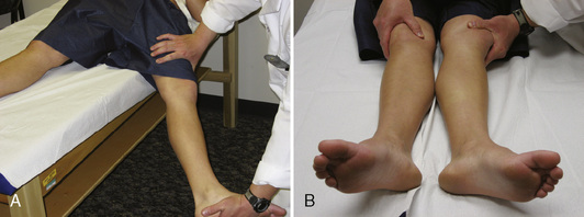
During the physical examination, the patient’s spine should first be examined to rule out other causes of hip pain. This should be followed by an examination of the elbows, hands, and knees to look for signs of hypermobility. Attention should also be paid to the patient’s skin and eyes. Next, the range of motion of both hips should be assessed. These patients often have an increased range of motion as a result of capsular laxity, but any significant increase in internal rotation should heighten one’s suspicion of increased femoral anteversion or other osseous abnormalities. This is followed by a thorough neurovascular examination that includes the reflexes. Next, specific hip testing should be performed, including the Ober test, the bicycle test, the psoas test, and the impingement test. In many cases, this process alters dynamic stabilizers (e.g., the iliopsoas) and leads to psoas and flexion contractures, internal coxa saltans, low back pain, and sacroiliac joint pain. Finally, hip-specific testing for capsular laxity should be performed. Patients with this condition will usually experience anterior hip pain while in the supine position with passive hip extension and external rotation (Figure 15-2, A). Patients may also have increased external rotation in full extension and distraction on the affected side (see Figure 15-2, B). Philippon and colleagues classified capsular laxity on the basis of these physical examination findings from grade 1 (mild) to grade 4 (severe), with grade 4 representing collagen vascular diseases.


Imaging and diagnostic studies


The radiologic workup begins with plain radiographs, including an anteroposterior view of the pelvis, a weight-bearing anteroposterior view, and a cross-table lateral view of the affected hip. Additional studies that may be necessary include Judet oblique films to further assess the acetabulum. False-profile views are used to assess for dysplasia, and computed tomography scans with spot views of the distal femoral epicondyles can be used to assess for acetabular and femoral anteversion. Various radiographic indices have been described to assess osseous undercoverage; these include but are not limited to the Tönnis angle, the center-edge angle of Wiberg, the anterior center-edge angle of Lequesne and de Seze, the femoral head extrusion, and the subluxation index. Acetabular version can also be estimated on radiographs by assessing the relationship of the anterior and posterior walls. A crossover sign and a prominent ischial spine both represent a retroverted acetabulum. The degree of retroversion can be estimated by the location of the crossover of the anterior wall, with more inferior crossing suggesting increased retroversion.


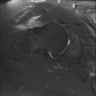
Magnetic resonance imaging (MRI) is critical for the evaluation of atraumatic instability. In the acute setting of traumatic hip injuries, numerous studies have demonstrated that MRI may aid in the diagnosis of chondral injuries, loose bodies, labral tears, femoral head contusions, sciatic nerve injuries, and ligament disruptions. Likewise, in the setting of chronic atraumatic injuries, MRI is also very useful to find subtle derangements in capsulolabral structures (Figure 15-3) as well as osteonecrosis.


Indications


Capsular laxity and atraumatic instability are difficult entities to assess, define, and ultimately treat; their management is still being developed. With the advent of better diagnostic and therapeutic capabilities, it is becoming increasingly clear that these pathologies exist. If a patient has a physical examination and history that are consistent with capsulolabral injury and instability and if appropriate imaging studies corroborate the clinical suspicion, then a trial of physical therapy and anti-inflammatory medication may be appropriately administered in an attempt to break the cycle of painful capsulolabral pathology. If conservative management fails and the patient has significant pain relief after an intra-articular anesthetic injection, then hip arthroscopy can be considered; however, because of the dearth of literature and unclear outcomes of this therapy, the mainstay of treatment for capsular laxity should still be conservative.


The anatomic restoration of the labrum (i.e., labral repair) and a reduction in capsular laxity either by capsular plication or thermal capsulorrhaphy have been described, with favorable preliminary results. In most cases, we perform capsular plication, because it is a reliable and measured reduction in capsular volume that is similar to the treatment of the shoulder. On rare occasions, when there is minimal capsular redundancy, we perform a thermal capsulorrhaphy; however, in most cases, we use the thermal device primarily as an adjunct. For cases in which atraumatic instability is a result of poor osseous coverage (e.g., acetabular dysplasia, excessive femoral valgus, increased femoral anteversion), hip arthroscopy should be approached with extreme caution, and redirectional osteotomies should be considered.


Surgical technique


The arthroscopic technique begins with adequate and appropriate anesthesia. Most often a general anesthetic is used with muscle relaxation, but regional anesthesia is also a possibility. With the patient supine on the operating room table, an examination under anesthesia is performed. Internal and external rotation is noted in full extension and in flexion and then compared with the nonoperative side. A flexion, abduction, and external rotation (FABER) test is then performed, with the distance from the lateral side of the affected knee being to the top of the operative table. As with external rotation, the affected limb often exhibits an increased amount of external rotation. Next, the hip is distracted, and a subjective evaluation of the necessary force needed to distract the hip joint is performed. Finally, if there is any concern regarding frank anterior instability, the limb is placed in extension, abduction, and external rotation, and a fluoroscopic image is performed to document this.


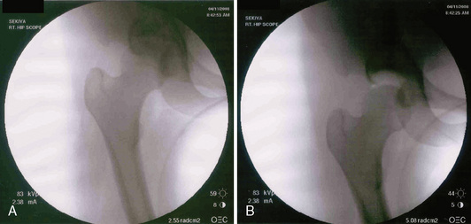
At this point, both of the patient’s feet are well padded, and the patient’s extremities are well secured to the fracture table. A well-padded perineal post is placed in between the patient’s extremities. The nonoperative limb is placed in full extension and mild abduction and then in minimal traction. The operative limb is put through a traction maneuver, which consists of abduction around the perineal post, axial traction, and adduction. Appropriate traction is then confirmed fluoroscopically. In cases of capsular laxity, minimal traction is usually needed to adequately distract the joint (Figure 15-4). The limb is then placed in internal rotation, which decreases the amount of hip distraction, reduces femoral anteversion, and subluxes the femoral head anteriorly, which enables the easy instrumentation of the joint. At this point, traction time is noted and documented.


< div class='tao-gold-member'>

Stay updated, free articles. Join our Telegram channel

Jul 24, 2016 | Posted by in MUSCULOSKELETAL MEDICINE | Comments Off on Arthroscopic Capsular Plication and Thermal Capsulorrhaphy

Full access? Get Clinical Tree

Get Clinical Tree app for offline access