Cervical spondylotic myelopathy (CSM) is a slowly progressive disease resulting from age-related degenerative changes in the spine that can lead to spinal cord dysfunction and significant functional disability. The degenerative changes and abnormal motion lead to vertebral body subluxation, osteophyte formation, ligamentum flavum hypertrophy, and spinal canal narrowing. Repetitive movement during normal cervical motion may result in microtrauma to the spinal cord. Disease extent and location dictate the choice of surgical approach. Anterior spinal decompression and instrumented fusion is successful in preventing CSM progression and has been shown to result in functional improvement in most patients.
Cervical spondylotic myelopathy (CSM) is a slowly progressive process that can be summarized as a gradual, progressive worsening of symptoms and a decline in functional status. In 1952, Brain and colleagues reported the first large series of patients with CSM. CSM is a result of the reduction in the effective space available for the spinal cord. The resultant changes in the spinal segments may compromise the integrity of the spinal cord secondary to static, dynamic, or ischemic mechanisms. CSM is the most common type of spinal cord dysfunction affecting individuals more than 55 years of age. Surgical treatment remains the most reliable and predictable method of preventing further neurologic deterioration.
Surgical rationale and decision making
The primary treatment of patients diagnosed with CSM is surgical. Nonoperative treatment consisting of medication, traction, or cervical collars has not been shown to alter the natural course of the disease. Up to 50% of patients treated conservatively deteriorate neurologically over time. Clarke and Robinson reported that 5% of patients deteriorate quickly, 20% have a gradual but steady decline in function, and 70% have a stepwise progression in their symptoms. Surgical intervention within 1 year of onset of symptoms can alter the natural history and change the prognosis of patients with CSM.
There are multiple surgical approaches to treating patients with CSM. The primary goal of surgery is to increase the canal diameter and thus to decrease the risk of both static and dynamic compression. This approach arrests progression, and, if done early enough in the disease process, can improve neurologic function. There are multiple factors that must be taken into account when considering either anterior or posterior decompression.
Applied surgical anatomy
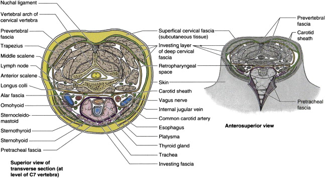
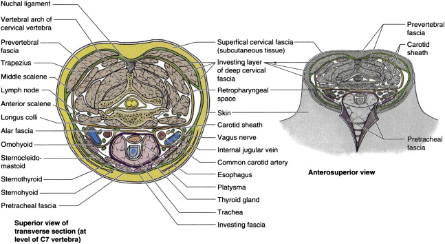
Choice of approach is dictated by the location of the disorder. The sternocleidomastoid (SCM), the carotid sheath, and the longus coli muscle serve as landmarks that differentiate the various approaches to the cervical spine. For example, approach can be either medial or lateral to the sternocleidomastoid. The key to the anterior cervical approach is in understanding the relationship between the neural structures, the vascular structures, the musculovisceral column and the 3 layers of the cervical fascia ( Fig. 1 ).

At-risk structures
Cervical Sympathetic Chain
The cervical sympathetic chain is located posteromedial to the carotid sheath and runs over the longus muscles. It extends longitudinally from longus capitis to longus colli over the muscles and under the prevertebral fascia. From superior to inferior, longus colli muscle diverges laterally, whereas the cervical sympathetic chain converges medially at C6. The average distance between the cervical sympathetic chain and medial border of the longus colli muscle at C6 is 11.6 (±1.6) mm. The superior ganglion of the cervical sympathetic chain in all dissections was located at the level of the C4 vertebra.
Vertebral Artery
The vertebral artery originates from the subclavian or innominate artery and enters the transverse foramen (TF) of the sixth cervical vertebrae. Before this, it is anterior to the transverse foraminae of C7. The artery passes through a series of foraminae until it reaches the base of the axis. It then turns posteriorly to enter the foramen transversarium in the posterolateral part of the ring of atlas and perforates the posterior atlantoaxial membrane to enter the foramen magnum. It then joins the contralateral vertebral artery to form the basilar artery, which supplies the brain stem and cerebellum.
The vertebral artery is intimately associated with the lateral border of the uncovertebral joint. Pait and colleagues reported that the distance from the tip of the uncinate process to the vertebral artery averaged 0.8 mm at C2 to C3 and 1.6 mm at C4 to C5. In a magnetic resonance imaging (MRI) study, Eskander and colleagues reported that the smallest distance between the uncovertebral joint (measured from the medial aspect of the uncovertebral joint to the medial aspect of the vertebral artery) was 4.15 mm at C3. Caution should be exercised when the uncinate process is removed in an attempt to remove osteophytes. In a review of 10 cases of vertebral artery injury, Smith and colleagues found that the use of a high-powered drill laterally was the most frequent cause of arterial injury. The surgeon must always check preoperative imaging for vertebral artery anomalies. There are 3 types of vertebral artery anomalies: (1) intraforaminal anomalies, (2) extraforaminal anomalies, and (3) arterial anomalies.
Recurrent Laryngeal Nerve
Injury to the recurrent laryngeal nerve (RLN) is a commonly reported complication following anterior cervical spine surgery. The RLN innervates all the intrinsic muscles of the neck with the exception of the cricothyroid. Almost all RLN injuries resolve over time; however, permanent paralysis rates have been reported in up to 3.5% of patients. Several causes for RLN injury have been suggested, including postoperative edema, stretch-induced neuropraxia, direct nerve ligature, direct retractor-induced nerve trauma, and nerve impingement between the endotracheal tube and retractor.
The right RLN branches from the vagus nerve at the level of T1 to T2. After looping around the subclavian artery, the right RLN becomes invested in the tracheoesophageal fascia (TEF). The RLN then travels superiorly and slightly anterior to the tracheoesophageal groove (TEG), before coursing between the trachea and the thyroid. In 82% (9 of 11) of right-sided dissections, the RLN entered the larynx at or inferior to C6 to C7. After looping around the aortic arch, the left RLN becomes invested in the TEF inferior to the T2 level. The nerve then travels slightly anterior to the TEG and within the TEF before coursing between the trachea and thyroid. In all the left-sided dissections, the RLN entered the larynx at or inferior to C6 to C7. The investigators concluded that both the left and the right RLNs had similar anatomic courses and received similar protection via surrounding soft tissue structures. From an anatomic perspective, the investigators did not appreciate a side-to-side difference superior to this level that could place either nerve at greater risk for injury.
Carotid Sheath
The carotid sheath contains the common and internal carotid arteries, the internal jugular vein, the vagus nerve, deep cervical lymph nodes, carotid sinus nerve, and sympathetic nerve fibers. The carotid sheath is a fascial investment that blends anteriorly with the investing and pretracheal layers of fascia. During surgical dissection, the carotid sheath is protected by the anterior border of the SCM. Care must be taken when placing and removing retractors to avoid vascular injury.
Esophagus
The esophagus is a thin ribbonlike structure that is located anterior to the retropharyngeal space. To prevent injury, it is critical to identify the esophagus during the approach. Placing an orogastric tube preoperatively can aide in esophageal identification.
Surgical considerations
Sagittal Alignment
Cervical sagittal alignment is an important consideration, because it affects the surgical treatment options and approach. The normal range of cervical lordosis is 40° (±9.7°) with most occurring between C1 and C2 while the occipital cervical junction is in kyphosis. The average lordosis between C4 and C7 is 6°, making this one of the flatter parts of the cervical spine. The presence of a cervical kyphosis poses a challenge for posteriorly based procedures. Because cervical laminectomy or laminoplasty are dependant on the posterior drift of the spinal cord, they are less effective in the kyphotic spine.
Restoring sagittal alignment should also be one of the goals of surgery and this may be more difficult to achieve from a posterior-only approach. Some of these difficulties may be overcome with an anterior approach or a combination anterior-posterior approach.
Disease Type and Location
The type and location of spinal cord compression dictate the surgical approach. The compression can be either soft or hard. Soft compression results from noncalcified soft tissues, whereas calcified or hard compressive disorders need to be identified because their presence may alter the surgical approach used to achieve decompression. Ventrally located compressive disorders can be localized to the level of the disk, dorsal to the vertebral body, or both. It can involve 1 or multiple levels. The addition of a dorsally located compressive disorder increases the complexity of decision making. Simple ventrally localized degenerative changes, such as anterior osteophytes or disk herniations, should be approached anteriorly to ensure adequate decompression. Dorsal compression, such as from ligamentum flavum hypertrophy or multilevel ossification of the posterior longitudinal ligament, should be approached posteriorly to achieve direct cord decompression. Sagittal alignment needs to be verified with a standing lateral radiograph. Because dorsal compression tends to be more generalized than focal, a posterior approach can more easily decompress multiple levels than anterior decompression. Generally, 1-level to 3-level ventral compressive disorders can be addressed from an anterior-only approach, whereas 4-level disorders should be addressed from a posterior approach or a combination anterior-posterior approach ( Fig. 2 ).

Motion Preservation
The incidence of adjacent segment degeneration following anterior cervical fusion has been reported to be approximately 3% per year. Motion preservation technology has gained popularity because it is intended to reduce abnormal stresses transferred to adjacent levels, thus theoretically reducing the chance of developing adjacent segment degeneration. Clinically, the risk of developing adjacent segment degeneration has been shown to be equivalent to both fusion and arthroplasty. There is also a concern that motion preservation may continue to result in microtrauma to the spinal cord, thus negatively affecting clinical outcomes. Buchowski and colleagues reported that 2 years after surgery, cervical arthroplasty outcomes were equivalent to arthrodesis for the treatment of cervical myelopathy for a single-level abnormality localized to the disk space. Hybrid surgery in which 1 level is fused and the second level is treated with cervical arthroplasty has been reported to be superior to 2-level anterior cervical diskectomy and fusion (ACDF) in better Neck Disability Index recovery, lower postoperative neck pain, faster C2 to C7 range of motion (ROM) recovery, and less adjacent ROM increase. Cervical arthroplasty is an option for the treatment of uncomplicated CSM. Whether adjacent level degeneration is the result of the fusion, or simply the expression of the natural deterioration of the motion segments, is unclear.
Surgical options
Corpectomy Versus ACDF for 2-Level Disease
When dealing with multilevel disease, the surgeon has to decide whether to perform an ACDF, a corpectomy, or a hybrid procedure ( Fig. 3 ). Both have unique advantages and disadvantages. The location of the disorder plays a dominant role in the decision-making process. Compression localized to the level of the disk can be approached with either procedure, whereas a disorder localized dorsal to the vertebral body needs to be treated with a corpectomy. The main advantage of a corpectomy in treating a multilevel disorder is the decreased number of graft-bone surfaces that need to fuse. In a 2-level ACDF, there are 4 bone-graft interfaces, compared with only 2 for a corpectomy. The theoretic risk of pseudarthrosis is therefore generally less with corpectomy than with multilevel ACDF. In a recent meta-analysis Fraser and Härtl reported that, for 2-disk–level disease, there was no significant difference between anterior cervical diskectomy with a plate system and corpectomy with a plate system. However, for 3-disk–level disease, the investigators reported that corpectomy with plate placement was associated with higher fusion rates than diskectomy with plate placement. Park and colleagues compared 2-level diskectomy with single-level corpectomy and reported comparable results for sagittal alignment, cervical lordosis, graft subsidence, and adjacent level ossification. Wang and colleagues compared 32 patients undergoing a 2-level ACDF with autograft with 20 patients with a 1-level corpectomy and anterior instrumentation. The investigators reported similar fusion and complication rates between the 2 groups. In contrast with these studies, Guo and colleagues reported that ACDF was superior in most outcomes to anterior cervical corpectomy and fusion and anterior cervical hybrid decompression and fusion. The investigators concluded that, if the compressive disorder could be resolved by diskectomy, then an ACDF is the treatment of choice.

ACDF
ACDF is commonly used in the treatment of CSM. This approach allows for spinal cord and nerve root decompression secondary to herniated disks and spondylotic bone spurs. With the use of an operating microscope, excellent visualization of the disk space, uncovertebral joints, and spinal cord is possible. A thorough diskectomy and meticulous end-plate preparation are critical for neural decompression, disk space distraction, and interbody fusion. This surgical approach uses intermuscular plane and is usually better tolerated than posterior surgery. Nerve root decompression can also be performed by resecting the uncovertebral spurs and foraminal decompression. Correction of sagittal plane deformities can be achieved by using lordotic intervertebral spacers. In addition, restoring anterior disk height increases the foraminal space and indirectly decompresses the nerve root. Single-level ACDF is associated with high fusion rates, particularly when used in conjunction with anterior plating. In a study of 2-level and 3-level ACDF with anterior plating, Samartidis and colleagues reported a fusion rate of 97.5% with either autograft or allograft. Wang and colleagues reported a high percentage of good to excellent clinical outcomes and a low incidence of segmental kyphosis and pseudarthrosis using anterior cervical plating.
Corpectomy
Cervical corpectomy is performed by removing the disk above and below the vertebral body to be resected. This removal is followed by resection of the vertebral body itself. Anterior osteophytes, ossification of the posterior longitudinal ligament (PLL), and disk herniations located behind the vertebral body can be addressed with this approach. Preoperative kyphosis can also be corrected. The procedure involves the creation of a trough in the middle portion of the vertebral body. It is critical to identify the lateral cortical wall of the vertebral body, which is located at the junction of the transverse process and the vertebral body. Identification of this landmark keeps the trough centered and symmetric, thereby avoiding injury to the vertebral artery. The wide trough permits extensive visualization of the disk, PLL, and osteophytes, thereby allowing a safe resection of all compressive disease. The strut graft for reconstruction following corpectomy is typically either structural iliac crest autograft, fibula allograft, or a cage construct packed with autograft. The incidence of surgical-related complications was associated with increasing patient age. Potential technical issues with these grafts include difficulty in restoring normal lordosis, graft dislodgment, graft migration, and, more commonly, graft subsidence. Anterior cervical plating is recommended for cervical corpectomy.
Multilevel Corpectomy
Multilevel disorder may require more complex surgical management. For corpectomy involving 3 or more levels, an anterior-posterior fusion has been recommended. Stand-alone anterior constructs have a high rate of graft migration, dislodgment, and pseudarthrosis. Construct motion has been shown to be lowest in a combined anterior-posterior instrumentation model. Circumferential cervical fusion has been shown to maximize cervical stability and decrease the incidence of graft migration or dislodgment. Accosta and colleagues reported that multilevel instrumented corpectomy offered a biomechanically stable fixation and allowed for significant correction of preexisting kyphosis. Supplemental posterior instrumentation was thought to limit delayed cage subsidence and loss of sagittal balance after this procedure. In the absence of posterior instrumentation, a large moment arm is created with an anterior-only construct. This construct can lead to loss of stability and pistoning or toggling of the graft and its eventual dislodgment, even with the use of an anterior plate or supplemental external fixation in a halo device. Vaccaro and colleagues treated 45 patients with 2-level or 3-level corpectomies and anterior plating. No supplemental posterior instrumentation was used. The investigators reported a 9% dislodgment rate in the 2-levelcorpectomy and 50% dislodgment rate in the 3-level corpectomy. Graft migration occurred despite the use of a halo in 10 of 12 patients who underwent a 3-level instrumented corpectomy. Sasso and colleagues reported a 71% catastrophic failure rate involving graft-plate dislodgment that occurred within 2 months of instrumented 3-level corpectomy. An additional advantage of supplemental posterior instrumented fusion is the ability to perform a posterior laminectomy, if needed, to further decompress the neural elements.
Stay updated, free articles. Join our Telegram channel

Full access? Get Clinical Tree


