Anatomical Landmarks for Therapeutic Massage

Chapter 2 Anatomical Landmarks for Therapeutic Massage


Knowledge of anatomy, especially that which is on the surface or creates superficial landmarks, is essential for performing therapeutic massage. It is important to know in which direction muscles run, where they attach to bone or cartilage, their relationships with blood vessels and nerves, and whether these structures are deep or superficial. Bony structures visible or palpable from the surface are key elements to identifying these soft tissue structures. Arteries and nerves are delicate and easily damaged, so it is vital to be aware of their position when performing soft tissue massage. Also, deep friction massage can be done to specific tendons and ligaments, so knowledge of their position is important.


When describing or defining the position of structures of the body, it is important to always maintain relationships with respect to the anatomical position. The anatomical position is defined as the body standing erect, head facing forward, trunk straight, and upper limbs extended with the palms upward. All structures are related to one another with the body in this position.


HEAD AND NECK


Therapeutic massage of the head and neck region must be done carefully. Several important nerves and blood vessels travel through this area, often superficially. Knowledge of these structures is important to avoid causing damage, either temporary or permanent.


Head


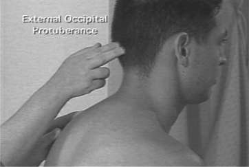
Bony prominences of the head and neck provide a frame of reference for location of important structures. The mastoid process is a prominent bony process of the temporal bone and is found posterior to the ear. It is important as the insertion for the sternocleidomastoid muscle. Another significant bony prominence is the external occipital protuberance (or inion), found on the occipital bone (Figure 2-1). Extending caudally from the external occipital protuberance and the posterior border of the foramen magnum, there is a superficial ligament of significance, the ligamentum nuche (or nuchal ligament). This structure extends from the external occipital protuberance along the spinous processes of the cervical vertebrae. This ligament helps to provide a site for muscle attachments.


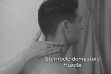
Laterally, another structure of significance in the head is the transverse process (TP) of the second cervical vertebra (C2). This landmark is identified by first locating the angle of the mandible. The TP of C2 is found between the angle of the mandible and the mastoid process. An important muscle landmark in the neck, just posterior to the TP of C2, is the sternocleidomastoid muscle (Figure 2-2), best viewed with the neck extended against resistance. This muscle originates from the manubrium of the sternum and the clavicle and inserts onto the mastoid process of the temporal bone and the superior nuchal line of the occipital bone.


Neck


Posterior to the sternocleidomastoid muscle is a large muscle of the neck, the trapezius. When the patient’s shoulders are elevated, this muscle is easily visible. This muscle extends from the skull and the ligamentum nuchae, along the spinous processes of cervical and thoracic vertebrae, to insert onto the clavicle as well as both the spine and acromion of the scapula.


Deep to the trapezius muscle are the splenius muscles (cervicis and capitis). These muscles reflect the meaning of the word splenion, meaning bandage, and extend from the spinous processes of cervical and thoracic vertebrae to the transverse processes of the cervical vertebrae and the skull. In the posterior neck, this muscle is found in a space bounded by the trapezius muscle posteriorly, the levator scapulae inferiorly, and the sternocleidomastoid muscle anteriorly.


Anterior to the splenius muscles are three pairs of scalene muscles: anterior, middle, and posterior. These muscles are involved in respiration and are important landmarks in the neck for locating the roots of the brachial plexus. The anterior and middle scalenes attach to the first rib, whereas the posterior scalene attaches to the second rib. All three muscles originate from the transverse processes of the fourth through sixth cervical vertebrae. At this origin, all three muscles can be palpated as one muscle mass, posterior to the sternocleidomastoid muscle. The roots of the brachial plexus are found in the neck between the anterior and middle scalenes.


UPPER LIMB


Massage to the upper limb most often involves the superficial muscle groups present. Although nerves and vessels are present in the upper limb, most are deep to muscle compartments and thereby protected. Massage to the upper limb is commonly needed in athletes, repetitive injury rehabilitation patients, or breast cancer patients.


Shoulder


Bony landmarks of significance in the shoulder are focused on the scapula and the clavicle. The clavicle is palpated on the anterior and superior aspect of the chest. Inferior to the clavicle is the space occupied by the subclavian artery, from which a pulse can be felt.


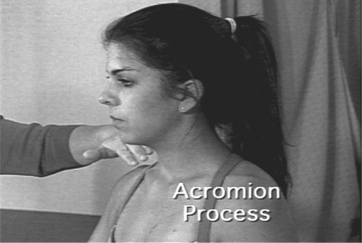
The scapula is a prominent flat bone on the dorsal aspect of the shoulder, between ribs 2 and 7. It articulates with the clavicle anteriorly and the humerus laterally. The medial border of the scapula can be pulled away from the back, along with the inferior border (at T8). The anterior border of the scapula is palpated best from the axilla. The most prominent posterior portion of the scapula is the spine (at T3), which provides a surface landmark to divide the posterior aspect of the scapula into supraspinous and infraspinous portions. The lateral end of the spine widens to form the acromion (Figure 2-3). At the angle of the acromion, where it changes direction, the deltoid muscle originates to form a cap over the shoulder. An additional important bony prominence on the scapula is the coracoid process. This process is medial to the head of the humerus as well as the acromion and is inferior to the clavicle. Several important muscles attach here (pectoralis minor, coracobrachialis, short head of the biceps).


The head of the humerus articulates with the scapula to form the shoulder, or glenohumeral joint. Just distal to the head of the humerus are the greater and lesser tubercles. By gripping the clavicle and the acromion and rotating the shoulder, the therapist can feel the head of the humerus as it rotates and can also feel the greater and lesser tubercles.


The deltoid muscle caps the shoulder. It has anterior, intermediate, and posterior portions. With the upper extremity in the anatomical position, the anterior portion is visible when the patient flexes the shoulder against resistance. The intermediate portion is visible when the patient abducts the arm against resistance. With the shoulder in extension, the posterior portion of the deltoid is visible at the shoulder.


Arm


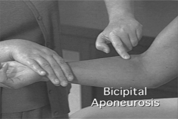
The arm, or brachium, is separated into anterior and posterior compartments. The most superficial, visible muscle of the anterior compartment is the biceps brachii muscle. It has, as the name implies, two heads of origin. The short head takes its origin from the coracoid process of the scapula, whereas the long head originates from the supraglenoid tubercle within the shoulder joint. Both heads are observed with the arm in the anatomical position. With the patient’s shoulder flexed and forearm supinated, the therapist can easily see the belly of the long head along its entire length until it disappears under the anterior portion of the deltoid. The insertion of the biceps, the biceps tendon, can be palpated as it attaches to the radius. The biceps muscle also ends as an aponeurosis (Figure 2-4), or broadened flat tendon, inserting into the ulna. This bicipital aponeurosis forms the roof of the cubital fossa and protects the brachial artery and the median nerve.


The triceps muscle, with three heads of origin, is the major muscle of the posterior compartment of the arm. The lateral head is located on the lateral aspect of the arm, the long head is medial and proximal, and the short head is distal.


The olecranon process is an easily palpated proximal posterior prominence of the ulna. It can be felt with the upper limb in extension, and, even more pronounced, with the elbow in flexion. The olecranon fossa, a depression in the distal humerus, can be palpated with the elbow in flexion. It is the site where the olecranon process of the ulna rests when the elbow is extended.


The medial and lateral epicondyles are bony prominences at the distal end of the humerus. The medial epicondyle is the site of attachment for many of the flexors of the forearm (pronator teres, flexor carpi radialis, palmaris longus, flexor carpi ulnaris, and flexor digitorum superficialis), as well as the landmark for locating the ulnar nerve as it passes posterior to the medial epicondyle. The ulnar nerve is very superficial, running in a groove posterior to the medial epicondyle of the humerus. It can be palpated both proximal and distal to this point, as a tight cord. The medial epicondyle feels sharp and is more prominent than the lateral epicondyle. It provides a point of attachment for many of the forearm extensor muscles (extensor carpi radialis brevis, extensor digitorum, extensor digiti minimi, extensor carpi ulnaris, and supinator).


Medial and lateral thickenings of the fibrous capsule of the elbow are the radial collateral (lateral) and ulnar collateral (medial) ligaments. The radial collateral ligament is fan shaped and runs from the lateral epicondyle of the humerus to blend with the annular ligament of the radius. The ulnar collateral ligament extends from the medial epicondyle to the coronoid and olecranon processes of the ulna. It is composed of three bands, although these are not distinguishable on palpation.


An important anatomical space on the anterior upper limb is the cubital fossa. This space is found anteriorly at the transition from the arm to the forearm. The cubital fossa is defined superiorly by a line connecting the medial and lateral epicondyles of the humerus. The inferior medial border is the pronator teres muscle, and the inferior lateral border is the brachioradialis. The cubital fossa is an important region because of the presence of several structures. Superficially, the median cubital vein is the most common site for venipuncture. Found deep in the cubital fossa, the brachial artery and median nerve are protected by the bicipital aponeurosis.


Forearm


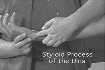
The ulna is the medial bone of the forearm and serves to stabilize this area. The posterior border of the ulna can be palpated, extending from the olecranon process to the wrist. The distal end of the ulna is the styloid process (Figure 2-5). This superficial bony eminence can be seen and palpated easily with the wrist in both extension and flexion.


The radius is the lateral bone of the forearm. It is shorter than the ulna. The head of the radius is a proximal structure found just distal to the lateral epicondyle. It is best palpated with the subject’s elbow flexed. Ask the subject to alternately supinate and pronate the forearm, and you will feel the radial head rotate as it articulates with the humeral capitulum. The groove felt between the radial head and the trochlea of the humerus during this rotation indicates the humero-radial joint line (HRJ). The radius ends distally as the styloid process. This process can be felt laterally, proximal to the thumb. The dorsal tubercle of the radius (Lister’s tubercle) can be palpated medial and posterior to the styloid process and separates the tendons of the extensor digitorum, extensor indicis, and extensor pollicis longus muscles from those of the extensor carpi radialis longus and brevis.


The wrist extensors are found in the posterior compartment of the forearm, and most originate as the common extensor tendon from the lateral epicondyle of the humerus. One prominent posterior compartment muscle, which is not a forearm extensor, is the brachioradialis muscle. The brachioradialis muscle is most obvious when the subject flexes the forearm against resistance, keeping the forearm in the neutral position. Once the brachioradialis muscle is identified, the rest of the superficial posterior compartment muscles can be determined. The extensor carpi radialis longus and brevis, extensor digitorum, and the extensor carpi ulnaris comprise the muscles in this compartment, from lateral to medial, relating to the brachioradialis. The tendons of these muscles, found on the dorsum of the wrist, are held in place during extension by the extensor retinaculum. The tendons of the extensor digitorum muscle are clearly visible on the dorsal surface of the hand, when the wrist is hyperextended.


Most of the forearm flexors originate from the medial epicondyle as the common flexor tendon. These muscles include the pronator teres, flexor carpi radialis, palmaris longus, flexor carpi ulnaris, and flexor digitorum superficialis. Placing your hand on the medial epicondyle and asking the subject to flex and ulnar deviate the wrist will cause the muscles originating here to be felt.

Stay updated, free articles. Join our Telegram channel

Jun 4, 2016 | Posted by in MANUAL THERAPIST | Comments Off on Anatomical Landmarks for Therapeutic Massage

Full access? Get Clinical Tree

Get Clinical Tree app for offline access