Acetabular Fractures: The Kocher-Langenbeck Approach


Acetabular Fractures: The Kocher-Langenbeck Approach


Berton R. Moed


INTRODUCTION

Fractures of the acetabulum are relatively uncommon injuries, usually resulting from high-energy trauma in young adult patients. Open anatomic reduction with internal fixation is the recommended treatment for fractures causing instability or incongruity of the hip joint. These fractures, which are often comminuted, are ideally treated by experienced fracture surgeons at institutions proficient in the care of multiply injured patients. Despite the best of care, however, these patients often have a protracted recovery and infrequently regain their preinjury level of physical capability (1).

These fractures constitute an anatomically diverse group of injuries. Judet, Judet, and Letournel (2) proposed the first systematic classification of acetabular fractures in 1961, which was based on the anatomic pattern of the fracture, and over time, this classification was modified and improved by Letournel. The comprehensive fracture classification systems of the Orthopaedic Trauma Association and the AO Foundation describe the alphanumeric coding of this “Letournel” classification and offer no clinical advantage. Therefore, the Letournel acetabular fracture classification continues to remain the international language of the majority of surgeons treating these complex injuries. This classification has 10 distinct categories, which are divided into five elementary types and five associated types (Fig. 41.1; Table 41.1).

The Kocher-Langenbeck, along with the extended iliofemoral and ilioinguinal, constitute the recommended “standard” approaches for the surgical treatment of acetabular fractures (2). Despite the advent of many alternatives, the Kocher-Langenbeck approach remains a mainstay in this regard (1).


INDICATIONS AND CONTRAINDICATIONS

As noted above, displaced fractures of the acetabulum resulting in joint incongruity or instability are best treated by open reduction and internal fixation (ORIF). Contraindications to surgery are ill-defined and not absolute. Important concerns include preexisting patient factors, such as poor general medical status and osteopenia, and factors that relate to overall patient prognosis, such as advanced age and associated injuries. All of these conditions must be considered with the knowledge that with nonoperative treatment in the face of joint incongruity or instability or both, the prognosis for hip-joint function is poor.

In choosing the appropriate surgical approach, the surgeon has an objective to select the least extensive exposure that allows sufficient bony access for anatomic joint reconstruction. The Kocher-Langenbeck approach provides direct visualization of the entire lateral aspect of the posterior column of the acetabulum (Fig. 41.2) (2). Indirect access to the true pelvis and to the anterior column can be attained by the palpating finger or through the use of special instruments (Figs. 41.2, 41.3 and 41.4) (2). Therefore, the Kocher-Langenbeck approach is applied in the treatment of fractures with the main displacement involving the posterior column. In the classification of Letournel (Table 41.1), this group consists of six fracture types: posterior wall, posterior column, posterior column plus wall, transverse, transverse plus posterior wall, and T-shaped. The Kocher-Langenbeck approach is the surgical exposure of choice for the first three types, in which the fracture extent is limited to the posterior wall or column or both. For the transverse, transverse plus posterior wall, and T-shaped fractures, some decision making is required. All three of these fracture types have a transverse fracture line as a common component. As a general guideline, if the fracture is <15 days old and the transverse component is located at (juxta-) or below (infra-) the level of the roof (tectum) of the acetabulum (therefore not involving the weight-bearing area of the acetabulum), the Kocher-Langenbeck approach is indicated (2). Otherwise, an alternative exposure, such as the extended iliofemoral approach, should be used. For acute juxtatectal- and infratectal-level transverse and T-shaped fractures, in which the major displacement occurs anteriorly at the pelvic brim and only minor posterior displacement, the ilioinguinal approach is perhaps the best choice.


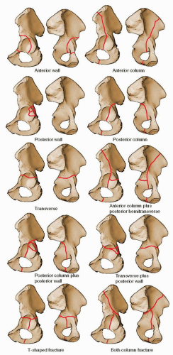
FIGURE 41.1 Letournel acetabular fracture classification. (From Moed BR, Reilly M. Fractures of the acetabulum. In: Rockwood and Green’s fractures in adults. 7th ed. Philadelphia, PA: Lippincott Williams & Wilkins; 2009:1475, Figure 45.14.)


TABLE 41.1 Acetabular Fracture Classification


Elementary fractures


Posterior wall


Posterior column


Anterior wall


Anterior column


Transverse


Associated fractures


Posterior column plus wall


Anterior column or wall plus posterior hemitransverse


Transverse plus posterior wall


T-shaped


Both column


From Letournel E, Judet R. Fractures of the acetabulum. Berlin: Springer-Verlag; 1981, and Letournel E, Judet R. Fractures of the acetabulum. 2nd ed. Berlin, Germany: Springer-Verlag; 1993.


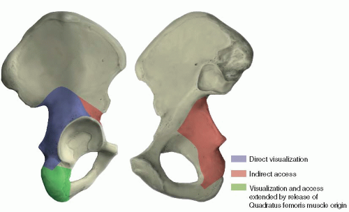
FIGURE 41.2 Access provided by the Kocher-Langenbeck approach.


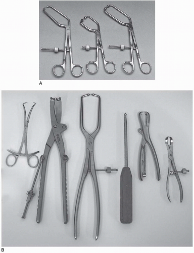
FIGURE 41.3 A,B. Examples of available instruments for acetabular fracture reduction. Special instruments that permit intrapelvic and anterior column access (A). Other useful reduction clamps, from left to right: large reduction forceps with points; pelvic reduction clamp; large pelvic reduction forceps with pointed ball tips; straight ball spike; Farabeuf reduction forceps; and serrated reduction forceps (B).

The status of the local soft tissues is an important additional consideration. Acetabular fracture surgery through a compromised soft-tissue envelope is ill-advised because of the increased risk of infection. Open wounds usually require débridement followed by delayed wound closure. Closed degloving soft-tissue injuries over the trochanteric region associated with underlying hematoma formation and fat necrosis (the Morel-Lavallee lesion) may be initially recognized by a fluid wave on palpation or may be later identified by the presence of a fluctuant, circumscribed area of cutaneous anesthesia, and ecchymosis. These injuries, even when closed, can be associated with the presence of pathogenic bacteria. Therefore, débridement followed by delayed wound closure and, subsequently, delayed fracture fixation may be required (2). This delay, as noted previously, may
preclude use of the Kocher-Langenbeck approach. More recently, a percutaneous method has been reported in a small number of patients, using a plastic brush to débride the injured fatty tissue, which is then washed from the wound with pulsed lavage (3). A medium closed-suction drain is placed within the lesion and removed when drainage is <30 mL over 24 hours. Fracture fixation is deferred until at least 24 hours after drain removal.


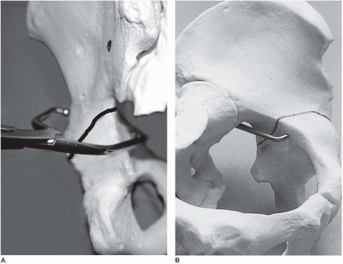
FIGURE 41.4 A,B. Example of clamp application for fracture reduction with a bone model. (A: Copyright Berton R. Moed, MD, St. Louis, MO, and Mark S. Vrahas, MD, Boston, MA. Permission granted.)


PREOPERATIVE PLANNING

In most cases, patients with an acetabulum fracture have sustained high-energy trauma. Therefore, examination of the injured limb, even in those with an apparent isolated injury, should be just one part of a comprehensive and systematic approach. Associated injuries can be life or limb threatening. The Advanced Trauma Life Support evaluation sequence should be followed (4). As previously noted, soft-tissue injury has important implications regarding subsequent surgery; therefore, the soft tissues should be evaluated carefully. The incidence of preoperative, posttraumatic, sciatic nerve injury was reported as being as high as 31% (5). Other peripheral nerves, such as the femoral and obturator nerves, also may be injured (6). A complete and clearly documented neurologic examination is extremely important both for patient prognosis and for medical-legal concerns. Preoperatively, this evaluation should be repeated periodically.

The initial anteroposterior (AP) x-ray of the pelvis can provide substantial diagnostic information regarding fracture type as well as indicate a need for emergency treatment (Fig. 41.5). This x-ray must be supplemented by further studies to define completely the acetabular fracture pattern. The three necessary additional plain x-rays (Fig. 41.6) are centered on the affected hip and include an AP and two 45-degree oblique views (the internal or obturator oblique view and the external or iliac oblique view) (2). Although these four plain x-rays usually provide all the information needed to define the acetabular fracture type, the standard two-dimensional computed tomography (CT) scan can supply important additional information and is indispensable for preoperative
planning (Fig. 41.7). The eventual universal availability of high-quality three-dimensional CT reconstructions may eliminate much of the mystery associated with the radiographic interpretation of acetabulum fractures (Fig. 41.8). However, except for the AP hip x-ray, which in most cases provides the same information as the AP pelvis examination, the plain and two-dimensional CT radiographic studies continue to be indispensable and should be viewed concurrently to make the definitive fracture diagnosis (2).


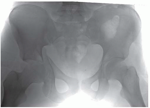
FIGURE 41.5 Initial AP pelvis x-ray of a 20-year-old man involved in a motor vehicle accident. There is a transverse fracture of the left acetabulum with a vertical fracture line through the ischium, suggestive of an atypical T-shaped pattern. A double density just lateral to the femoral head suggests a displaced intra-articular or wall-fracture component. The right hip is subluxed, but not dislocated, and there is widening of the right sacroiliac joint.

After careful physical examination and radiographic study, the appropriate surgical approach can be determined. The indications for emergency fracture fixation are uncommon (Table 41.2). Operative treatment is generally delayed 3 to 5 days to allow stabilization of the patient’s general status and for preoperative planning. My preference is to use preoperative, skeletal, femoral-pin traction both to maintain an unstable hip in a located position and to prevent further femoral head articular-surface damage from abrasion by the raw acetabular bony fracture surfaces (Fig. 41.9). Significant intraoperative blood loss can occur. Approximately 2 units of blood should be made available, depending on the extent of the fracture pattern. The use of an autologous blood transfusion system may decrease the need for intraoperative, homologous, banked-blood transfusion.


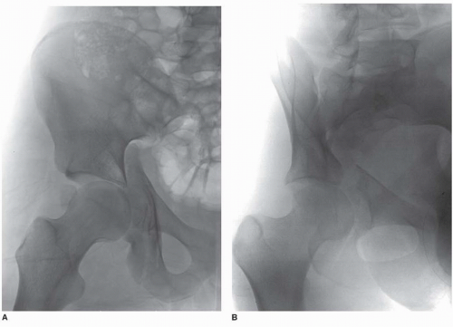
FIGURE 41.6 A-C. Subsequent AP and 45-degree oblique hip x-rays visualize the atypical T-shaped acetabular fracture more completely. However, the additional fracture components are not well delineated.


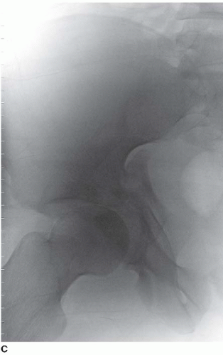
FIGURE 41.6 (Continued)


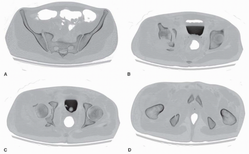
FIGURE 41.7 Selected two-dimensional CT sections. In addition to the previously noted findings consistent with an atypical T-type fracture, a posterior wall fracture fragment, and two osteochondral-free fragments (one intra-articular and one displaced anterior to the femoral head) are evident.


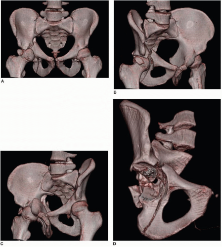
FIGURE 41.8 A-D. Three-dimensional CT constructs formed as AP and 45-degree oblique views (A-C) and created using the volume-rendering technique show very clearly the fracture as deduced by evaluation of the plain x-rays and two-dimensional CT. An excellent overall appreciation of the fracture pattern is provided. A three-dimensional construct subtracting the femur and oriented obliquely into the hip joint (D) shows the fracture comminution; however, there is some loss of definition.


TABLE 41.2 Indications for Emergency Acetabular Fracture Fixation


Recurrent hip dislocation after reduction despite traction


Modifier: None


Progressive sciatic nerve deficit after closed reduction


Modifier: None


Irreducible hip dislocation


Modifier: After open reduction (stable with traction), fracture fixation may be delayed because of declining medical status of the patient or limitations of the surgical team


Associated vascular injury requiring repair


Modifier: When the fracture is directly related to the vascular injury and fracture stabilization is an important adjuvant to the vascular repair, such as an anterior column fracture associated with laceration of the femoral artery, urgent fracture fixation is required


Open fractures


Modifier: Open fracture treatment principles require emergency irrigation, débridement, and fracture stabilization. Fracture stabilization options include traction followed by delayed ORIF or acute ORIF


From Tile M. Fractures of the pelvis and acetabulum. 2nd ed. Baltimore, MD: Williams & Wilkins; 1995, with permission.


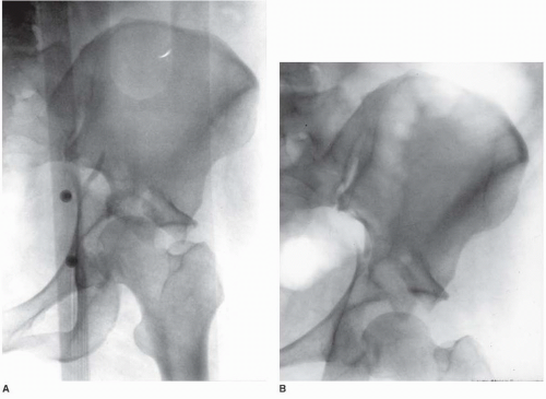
FIGURE 41.9 AP hip radiographs before and after the application of traction. A. Without traction, the femoral head is medially subluxed, rubbing against the sharp corner of the superior acetabular fracture surface in this displaced transtectal fracture. B. The hip joint is distracted with the application of traction pulling the articular cartilage of the femoral head a safe distance away from the acetabular fracture surface.


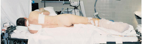
FIGURE 41.10 Lateral position for surgery on the right hip. The patient is supported on a beanbag on a radiolucent operating room table. The down leg is padded, and an axillary roll and head supports are in position. For this patient with an ipsilateral ankle fracture, the right leg is splinted and padded.


SURGICAL TECHNIQUE


Positioning

Acetabular fracture fixation using the Kocher-Langenbeck approach can be performed with the patient in either the lateral or the prone position. Orthopedic surgeons from North America are more familiar with and perhaps more comfortable using lateral positioning with the affected extremity draped free, as in hip arthroplasty surgery (Fig. 41.10). However, although definitive study is lacking, the Kocher-Langenbeck approach is generally thought to be most effective with the patient placed prone on a fracture table (1,7). The benefits of the prone position are realized by maintaining the femoral head in a reduced position. Gravity becomes a help rather than a hindrance in fracture exposure and reduction. The fracture table provides controlled traction and limb positioning, further assisting in fracture reduction. Traction is applied through use of a distal femoral pin with the knee flexed to approximately 90 degrees (Fig. 41.11). This angle of knee flexion places the sciatic nerve in a relaxed position, minimizing the risk of intraoperative sciatic-nerve injury. An unscrubbed assistant is required for intraoperative adjustment of the table.


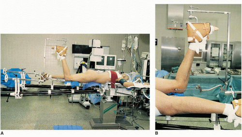
FIGURE 41.11 Patient in the prone position for surgery on the right hip (A) with a detailed view of the affected limb and femoral pin position (B).


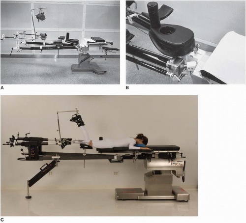
FIGURE 41.12 The Judet fracture table. A small pad can be used to elevate the patient’s head (A). A detailed view (B) shows the padded perineal post and the padded support with perineal cutout for male patients. The separation between the chest and padded perineal support serves to reduce abdominal pressure without requiring additional padding or chest rolls. The currently available PROfx Fracture Table version (C) manufactured by Mizuho OSI (Figure courtesy of Mizuho OSI, Union City, CA. Permission granted.)

With the patient placed prone, chest rolls should be used to elevate the head and to avoid excessive abdominal pressure. The fracture table generates the added risk of injury (i.e., pudendal nerve palsy) from pressure against the perineal post. The Judet fracture table adequately addresses these concerns. Although no longer being manufactured, the original, Tesserit T3000 model (Figs. 41.11 and 41.12) continues to be available in reconditioned form (Medrecon, Inc., Garwood, NJ). An updated version, which is also more practical for general fracture table usage, is currently available (Fig. 41.12C; PROfx Fracture Table, Mizuho OSI, Union City, CA).


C-Arm

No matter what the patient position, use of a radiolucent operating table is advisable. Intraoperative C-arm fluoroscopy can then be used to assess fracture reduction and hardware location (Fig. 41.13). Before the sterile preparation and draping of the patient, the hip area should be quickly scanned with the C-arm to ensure adequate fluoroscopic visualization.


FIGURE 41.13 Intraoperative fluoroscopic views of a transverse with an associated posterior-wall fracture before (A) and after (B) the transverse fracture component was reduced by using a pointed reduction forceps.


Draping

With the patient in the lateral position and the limb draped free, the sterile field is similar to that in hip arthroplasty surgery but extended posteriorly to include the region of the posterior-superior iliac spine (Fig. 41.14). With the patient prone on the fracture table, the sterile field consists of the buttock and the posterior and lateral aspects of the thigh (Fig. 41.15).


Surgical Approach

An overview of the surgical approach is shown in Figures 41.16A-C. The skin incision (Fig. 41.17) is centered over the greater trochanter. The proximal branch of the incision is directed toward the posterior-superior iliac spine, ending approximately 6 cm short of this bony landmark. Distally, the incision extends approximately 15 cm along the midlateral aspect of the thigh. This skin incision is carried through the subcutaneous tissue and superficial fascia onto the fascia lata of the lateral thigh (the iliotibial tract) and the thin, deep fascia overlying the gluteus maximus muscle (Fig. 41.18).

The fascia lata is then divided in line with the skin incision, beginning at the distal aspect of the wound, continuing proximally toward the greater tuberosity, and ending at the first sighting of the gluteus maximus muscle fibers as they insert into the iliotibial tract (Fig. 41.19). The trochanteric bursa of the gluteus maximus (a large bursa between the tendon of this muscle and the posterolateral surface of the greater trochanter) is incised, allowing clear visualization of the insertion area of the gluteus maximus muscle and access to the undersurface of this muscle (Figs. 41.20 and 41.21). Beginning the deep dissection in this way, at the distal branch of the Kocher-Langenbeck incision, the surgeon facilitates the next step: splitting of the gluteus maximus muscle.


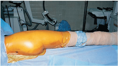
FIGURE 41.14 Patient from Figure 41.10 after sterile preparation and draping. The posteriorsuperior iliac spine is marked with an “X.” The right leg is draped free.

Only gold members can continue reading. Log In or Register to continue

Stay updated, free articles. Join our Telegram channel

Tags:
Aug 28, 2016 | Posted by in ORTHOPEDIC | Comments Off on Acetabular Fractures: The Kocher-Langenbeck Approach

Full access? Get Clinical Tree

Get Clinical Tree app for offline access