Abductor Muscle and Trochanteric Complications Following Total Hip Arthroplasty
Philipp Leucht
James I. Huddleston
Case Presentation
An otherwise healthy 76-year-old female undergoes a right primary total hip arthroplasty (THA) for advanced osteoarthritis. During her procedure, the treating surgeon recognizes a degenerative tear of the gluteus medius tendon at its insertion, which he treats with a primary repair during the index procedure. The patient tolerates the procedure well and recovers appropriately. The arthritic pain completely resolves, however, she is complaining of significant weakness in her operative hip and presents with an obvious Trendelenburg gait. Nine months after the index procedure, she still requires a cane for ambulation.
Introduction
Proper hip mechanics rely on balanced muscular support. Weakness of the abductor mechanism after THA can result in significant functional impairment for the patient. Restoration of the muscular balance of the hip joint can present a challenge for both the patient and the treating orthopedic surgeon. This chapter reviews the bony and soft tissue complications associated with fracture and osteotomy of the greater trochanter as well as sequelae from avulsions of the abductor mechanism. In addition, diagnostic algorithms, treatment options, and results of various techniques used to address these complications are discussed.
Fracture
Periprosthetic fractures of the greater trochanter can occur both intraoperatively or postoperatively. Intraoperative fractures during primary THA remain relatively common complications that necessitate appropriate recognition and management to prevent potentially significant sequelae. It is estimated that intraoperative, periprosthetic fractures occur in approximately 1% to 20% of primary THA cases (1,2,3,4,5,6). The incidence of isolated greater trochanter fractures ranges from 0.4% (7) to 5% (8). In a series of primary THAs performed through an anterior approach, the rate of intraoperative greater trochanter fracture was 0.7% (3/437 THAs) (9). If a fracture of the greater trochanter is recognized during the hip reconstruction, then repair should be attempted to avoid long-term problems such as trochanteric escape with resultant hip abductor weakness. Depending upon the size and shape of the fracture fragment, multiple surgical fixation options can be utilized. In cases where a small fragment is detached, but not significantly displaced, simple cerclage wiring with either metal of polyethylene cables, simple wires, or large-gauge nonabsorbable suture can be sufficient for stable fixation. As with all wire fixations, care should be taken to ensure close contact of the wire to bone during the passing, as interposed soft tissues significantly reduce fracture compression. To optimize the compressive force vector, the wire should be passed distal to the lesser trochanter and avoid contact of the wire with the prosthetic neck and thus, reduces the risk of fraying and subsequent particle generation within the effective joint space, which may in turn lead to clinically significant third-body wear at the hip articulation. In cases of substantial comminution of the greater trochanter and abductor insertion, a trochanteric cable grip system can provide a broader area of fixation and concomitant compression (Fig. 86.1). The postoperative regimen should include toe-touch weight bearing of the affected side for a minimum of 6 weeks or until radiographic healing is evident. Patients should be advised to avoid active hip abduction during this time. If there is any concern in regard to fracture fixation or patient compliance, a hip abduction orthosis should be considered. Once radiographic evidence of union is noted, patients can be slowly advanced to weight bearing as tolerated and active abduction exercises under guidance of a physical therapist.
The incidence of postoperative greater trochanter fractures is unknown. It has been estimated that postoperative periprosthetic femoral fractures occur in less than 1% of primary THAs (10). Postoperative greater trochanter fractures with less than 2 cm of displacement have been successfully
treated without operative intervention. Nonoperative treatment includes 6 to 12 weeks of limited weight bearing and no active abduction exercises until the fracture has united (8). Frequent radiographic follow-up is recommended to detect significant migration of the greater trochanter fragment. Despite a lack of scientific evidence, current recommendations suggest operative fixation of the greater trochanter fracture if the displacement exceeds 2 cm and the fragment is large enough to allow stable fixation (8).
treated without operative intervention. Nonoperative treatment includes 6 to 12 weeks of limited weight bearing and no active abduction exercises until the fracture has united (8). Frequent radiographic follow-up is recommended to detect significant migration of the greater trochanter fragment. Despite a lack of scientific evidence, current recommendations suggest operative fixation of the greater trochanter fracture if the displacement exceeds 2 cm and the fragment is large enough to allow stable fixation (8).
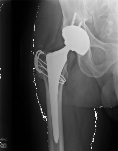 Figure 86.1. Radiograph of a trochanteric cable grip system. |
Nonunion of the Greater Trochanter
Nonunion of the greater trochanter, especially after osteotomy, is a common complication. Nonunion has been associated with the need for revision surgery, antalgic gait, pain, femoral loosening, and a lower Charnley Hip Score (11). Rates vary and depend on the method of fixation (12). First-generation techniques utilized stainless steel and cobalt-chrome alloy monofilament wires. This fixation technique demonstrated nonunion rates of 0% to 7.9% in studies that ranged in size from 75 to 1,162 patients (11,13,14,15,16,17,18). Risk factors for the development of nonunion after wire fixation alone include male gender, revision surgery, and a primary diagnosis of rheumatoid arthritis. Technical factors that may predispose to nonunion with this technique include small size of the osteotomy fragment, lack of sufficient wire tension, limited surgical experience, wires placed around the lesser trochanter, and apposition of the trochanter to a bed that is primarily cement (14,19,20). Studies have shown no association between the development of a nonunion and the type of osteotomy (uniplane vs. biplane) or the wire fixation technique (21,22). A meta-analysis of studies involving 2,910 THAs reported a wire breakage rate of 22% with various wiring techniques (13,14,15,20,21,23,24).
Multifilament cables were introduced in 1977 by Dall and Miles (25) as the second-generation fixation technique for greater trochanter osteotomies. Probably the greatest advantage of cables over wires is the greater strength of the cable and the ability to provide greater compression to the osteotomy site, while providing greater resistance to deformation (20,26,27,28,29,30,31,32,33). However, there are reports of initial cable relaxation of up to 50%, which can be detrimental for compression at the osteotomy site, resulting in a possibly higher nonunion rate (34). Despite improvement in mechanical properties, breakage rates for cables have been reported to be as high as 12% (20/160 hips) (24


Related posts:


Stay updated, free articles. Join our Telegram channel
Full access? Get Clinical Tree
