Procedure 29 Spondylolysis Repair
Indications
• Pars defect must be source of pain
• Minimal disk degeneration present
• Patient 30 years of age or younger
Examination/Imaging
• Anteroposterior and lateral lumbar radiographs
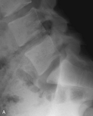
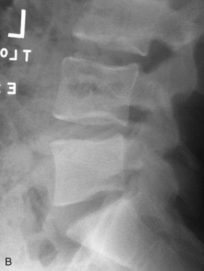
• Flexion and extension lateral lumbar radiographs (Figure 29-1, A and B)
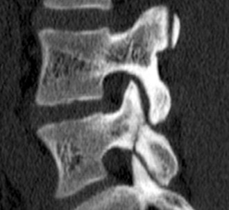
• Lumbosacral computed tomography (Figure 29-2)
Portals/Exposures
• A standard posterior lumbar exposure is used to access the affected vertebra. The lamina, facets, and proximal transverse process should be exposed (Buck, 1970; Askar, 2003; Chung, 2007; Debusscher, 2007).
• Recently, minimally invasive techniques of repair have been described using percutaneous screws and of tubular retractors (Nichol, 1986; Morscher, 1988).


Stay updated, free articles. Join our Telegram channel

Full access? Get Clinical Tree


