Fig. 5.1
Bony landmarks and ligament attachment sites. GT gastrocnemius tubercle, AT adductor tubercle, POL posterior oblique ligament, SMCL superficial medial collateral ligament, ME medial epicondyle, DMCL deep medial collateral ligament
The DMCL is a short and broad capsular thickening that attaches firmly to the meniscus and just beyond each articular margin of the tibia and femur. It functions as a secondary restraint to valgus load at all flexion angles.
The POL is a triangular thickening of the joint capsule with a firm attachment to the posterior horn of the medial meniscus. It is composed of three different fascial expansions: the superficial, central, and capsular arms. The central arm is the main restraint component, which is reconstructed in the presented technique. Its femoral attachment is posterior to the SMCL lying just distal (2.9 mm) to the gastrocnemius tubercle, an osseous prominence coined by LaPrade et al., which is a very helpful intraoperative identification [6]. It can also be located distal and posterior to the adductor tubercle. The tibial attachment is anterior to the semimembranosus tendon with an expansion to the anterior portion of the semimembranosus tendon allowing the POL to be loose in flexion and taught in extension, especially with internal rotation.
5.2 Clinical Relevance
Clinically, the medial ligaments provide approximately 57 % of restraint to valgus stress at 5° flexion [9]. Increasing the flexion angle increases load bearing on these structures yielding up to 78 % of restraint at 25° [9]. When the distal portion of the SMCL is ruptured, studies have shown there to be just as much external rotation instability as found in posterior lateral corner disruptions [8].
There is however debate to the proper treatment of MCL complex injuries partly in due to the fact that the SMCL has served as a model for ligament healing in basic research. The SMCL has a large reserve for healing nonoperatively and is extrasynovial, unlike the ACL, which has poor healing reserve and typically requires surgical reconstruction. The healing process of the MCL has been well studied and consists of the inflammation phase (72 h), repair and regeneration phase (6 weeks), and the remodeling phase (can be present up to 1 year) [10].
Knee laxity is graded by medial joint opening with valgus force at 30°: 0 (normal), I (1–4 mm), II (5–9 mm), and III (10–15 mm). Grades I and II will have a hard endpoint, while grade II injuries will have a soft endpoint as the MCL is considered fully ruptured at this grade. Generally the preferred treatment for isolated acute MCL complex injury is nonoperative management. Grade I and II laxity require hinged bracing and guarded weight bearing initially followed by early range of motion. Nonoperative management has been shown to provide good patient-reported outcomes and even grade III type lesions go on to heal regularly with patients returning to full activity levels in 5–7 weeks time [7, 11–13].
However, studies have shown that grade III injuries often heal with less stability than the uninjured side [14]. Animal studies have demonstrated an increase in type III collagen scar following healing, resulting in increased laxity and only 70 % of initial strength [15–17]. Other studies have noted improved strength with surgical repair and improved results and strength with early nonoperative range of motion [2, 11, 18]. Propensity to heal is highly correlated with the location of the rupture with mid-substance tears reserving the most potential for healing, less so as one moves toward the bony attachments [19, 20].
5.3 Presentation
Patients with acute injuries usually present following a traumatic event with localized swelling and induration over some portion of the MCL location. Most low-grade sprains occur as noncontact injuries during a valgus and external rotation load. Complete MCL disruptions often result from a direct blow to the lateral leg. These pure valgus forces result in an isolated MCL injury. The addition of external rotation forces to the tibia expands the zone of injury to include the POL and/or the anterior cruciate ligament. As 78 % of all grade III injuries are multiligamentous, an audible pop and generalized intra-articular swelling might suggest anterior cruciate involvement [12]. Although often confounding in the acute patient, deep medial joint line tenderness can be indicative for medial meniscal injury.
Chronically MCL deficient patients will typically describe mostly side-to-side instability and pain. These patients tend to present after minor injury or activity aggravating their instability. The Swain test, described by Lonergan and Taylor, is a testing for chronic deficiency and rotatory instability [22]. Performed with the knee flexed to 90°, the tibia is then externally rotated. In this position the cruciate ligaments are lax while the collaterals are taught. Medial pain will often be present with chronic laxity or inadequate ligament healing. While in this position, one can perform anteromedial drawer test to evaluate POL integrity [5]. Valgus gapping at 30° flexion will evaluate SMCL stability. Valgus gapping with the knee in full extension is more indicative of a combined SMCL and POL injury [23]. Of note, patients with a POL injury may also exhibit a positive Dial test, as anterior rotatory instability due to deficient POL will confound this test [5]. All grade III tears should undergo examination for concomitant cruciate injury, as mentioned above most are not isolated MCL injuries. Any evidence of genu valgum should be evaluated and treated before attempts are made to reconstruct medial ligaments.
Care should be taken to obtain high-quality radiographs on all patients. In addition to evaluation of fractures, avulsions, and loose bodies, radiographs should be obtained to delineate previous hardware and tunnel placement, osteophyte formation mechanical alignment, and lateral capsular signs (Segond’s fracture); Pellegrini-Stieda lesions are specific for chronic MCL lesions. Stress views to evaluate gapping, and physeal injuries in adolescents should also be obtained.
MRI can be most useful when coupled with the physical exam to delineate the location of MCL tears, meniscal tears, cartilage lesions, and associated cruciate lesions.
5.4 Surgical Indications and Contraindications
While indications for surgical reconstruction of MCL complex injuries still remain highly debated, the set of indications the senior author follow are listed in Table 5.1.
Table 5.1
Surgical indications and contraindications
Indications | Bony avulsion injury |
Chronic instability | |
Continued instability beyond 6 weeks of nonoperative management | |
Failed ACL repair with medial instability | |
Contraindications | Isolated grade I or II injuries |
Skin disruption or breakdown over medial skin |
5.5 Technique
In a subset of the acutely injured, operative repair of the effected medial ligaments is indicated. In patients who are initially treated by nonoperative means but continue to show valgus instability and are clinically symptomatic, reconstruction is indicated. It has been the experience of the senior author that optimal function of the MCL complex deficient patient is restored through reconstruction that reestablishes the patient’s native anatomy. The technique of the senior author involves a reconstruction of the proximal and distal divisions of the SMCL and POL using two separate grafts.
The patient is placed supine on the surgical bed. General anesthesia is used. A thigh tourniquet is applied to the operative leg. The foot of the bed is flexed maximally with enough room behind the flexed knee to allow a fist to be placed in times when manipulation is required during the procedure. Two rolled sheets placed under the pad of the bed prevent hyperextension at the hip. The contralateral lower extremity is safely cradled in a well-type leg holder with the fibular head well padded. The nonoperative leg is also fixed with the knee and hip slightly flexed and abducted (Fig. 5.2).
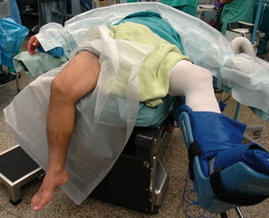
Fig. 5.2
 Acute Lateral and Posterolateral Injury
 Surgical Approach to Posterolateral Chronic Injury
 Postoperative Management: Rehabilitation
 Treatment for Chronic Knee Dislocation
 Chronic Lateral and Posterolateral Laxity: Reconstruction Techniques
 Chronic Anterolateral Knee Laxity: Reconstruction Techniques


Patient position and setup before sterile draping
Related posts:
Acute Lateral and Posterolateral Injury
Surgical Approach to Posterolateral Chronic Injury
Postoperative Management: Rehabilitation
Treatment for Chronic Knee Dislocation
Chronic Lateral and Posterolateral Laxity: Reconstruction Techniques
Chronic Anterolateral Knee Laxity: Reconstruction Techniques
Stay updated, free articles. Join our Telegram channel
Full access? Get Clinical Tree
