Fig. 47.1
Simple bone cyst in its most common site. Well outlined centrally and symmetrically located metaphysiodiaphyseal lucency. The cortices are expanded and thinned. The lesion is not wider than the epiphyseal plate. Cysts located near the plate are called “active”
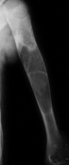
Fig. 47.2
Simple bone cyst located in the diaphysis of humerus. In long bones, growth may result in the cyst moving away from the plate and finishing located in the diaphysis. This kind of cyst is called “latent” or “inactive”
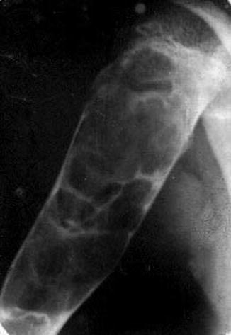
Fig. 47.3
SBC showing a multilocular appearance due to prominent endosteal bony ridges in the inner cortical wall
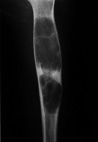
Fig. 47.4
A diaphyseal location in humerus. The sclerotic area is probably the result of a previous fracture
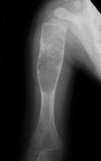
Fig. 47.5
“Active” SBC with a large extension to the diaphysis. The cyst shows a conic end in the shaft, with a sharp margin with trabeculation
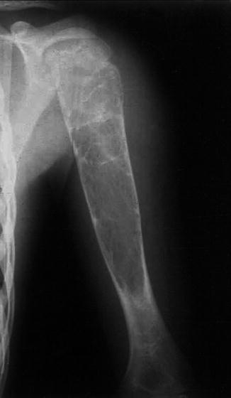
Fig. 47.6
SBC with trabeculations in the metaphyseal area and a prominent extension in diaphysis
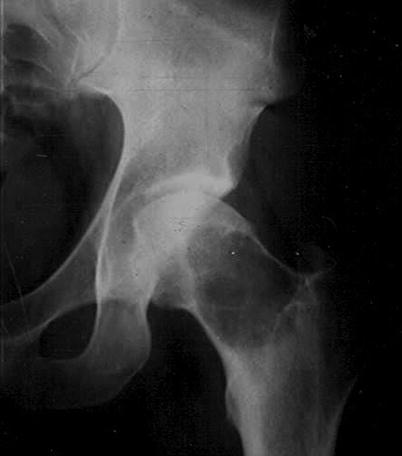
Fig. 47.7
SBC in the second most common site, the upper end of femur. This cyst is located in the femoral neck
Fig. 47.8
(a) SBC located in the intertrochanteric area of the upper end of femur. Periosteal reaction is always absent in SBC except at the sites of fracture. (b) MRI usually confirms the cystic unicameral nature and its fluid content. (c) CT scan, well-delimited lesion that thinned the cortex. True septation is unusual and may be present after fractures
Fig. 47.9
(a and b) Two patients with solitary bone cyst affecting the upper end of the femur. In the two cases the cysts present well-delimited margins and trabeculations or pseudoseptations. The lesions are centrally located and do not expand the cortex
Fig. 47.10
(a, b) Anteroposterior and lateral X-ray showing an SBC of the upper end of the tibia, with an eccentrical location
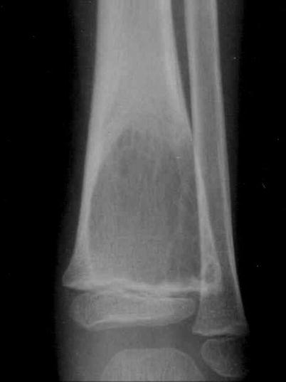
Fig. 47.11
SBC in distal metaphysis of a tibia, adjacent to the physis
Fig. 47.12
(a) SBC in the ilium. This location is affected more often in older patients. (b) CT scan shows a lytic lesion showing a pseudoseptation. (c) MRI shows a cystic unicameral lesion with fluid content
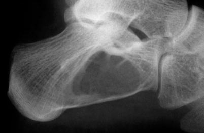
Fig. 47.13
Typical aspect of an SBC arising in the calcaneus. In this site lipoma of bone is the most common image differential diagnosis
Fig. 47.14
(a, b) Typical roentgenogram and CT of an SBC of the calcaneus
Fig. 47.15


(a–c) SBC arising in the calcaneus with typical MRI pattern in T1- and T2-weighted images
Related posts:


Stay updated, free articles. Join our Telegram channel
Full access? Get Clinical Tree
