Fig. 9.1
Types of FAI
Acetabular Deformity
An unusually deep acetabulum can cause FAI because the femoral neck is excessively covered. Alternatively a retroverted acetabulum will cause impingement against the femoral neck during hip flexion and abduction.
Acetabular Dysplasia Classification
- A.
Classical: Classical DDH involves reduced coverage of the femoral head and is therefore not usually a type of dysplasia that contributes to FAI, but has been included for completion.
- a.
Deficient head coverage
- b.
Decreased acetabular depth
- a.
- B.
Acetabular retroversion: anterior wall is rotated forward or posterior wall is rotated backward
- C.
Acetabular over coverage: anterior and lateral wall over coverage of the femoral head.
Femoral Head Deformity
This can take two forms. Pistol grip deformity occurs when the femoral head loses sphericity and the head neck junction is less pronounced. The appearance is likened the handle of a pistol as shown in Fig. 9.2.
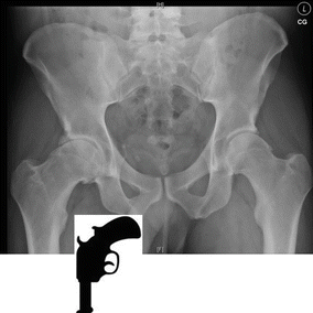
Fig. 9.2
Diagram of pistol grip deformity
The femoral head can also develop a tilt deformity that is apparent superomedial migration of the femoral head.
Femoral Neck Deformity
This involves a shortened or widened femoral neck, which reduces the head-neck ratio.
What Are the Predisposing Factors for FAI?
Epidemiology
Males are affected more commonly than females. Men are more likely to have cam deformities and women are more likely to have acetabular retroversion or protrusio causing pincer type FAI. The typical age range is 14–45.
Childhood hip disorders such as slipped upper femoral epiphysis (SUFE), developmental hip dysplasia (DDH) and Perthes’ disease can lead to secondary FAI.
SUFE is the leading cause of FAI contributing to its development in up to 50 % of cases [3]. Any degree of displacement of the capital femoral epiphysis can cause the femoral head to lose its spherical shape or affect the femoral head-neck ratio; however, the most common deformity caused by SUFE is a cam deformity. FAI can occur whether SUFE has been diagnosed and managed appropriately or missed. Even mild cases of SUFE that have been treated with percutaneous pinning show FAI changes on imaging in around 50 % of patients [4].
DDH can lead to FAI due to the potential for permanent deformity of the femoral head, acetabulum or head-neck shape. This can occur whether DDH is missed or managed with non-operatively or operatively. Following femoral varus osteotomy or pelvic osteotomy, it is possible for iatrogenic FAI to occur. Similarly, the incongruity and cam deformity associated with Perthes’ can cause FAI and OA in later life.
Sporting activities that force the hip through repetitive or a high range of movement can cause impingement at the extremes of movement. Specific sports associated with FAI include gymnastics, dance, hockey and football. Elite athletes have a higher incidence of FAI than the general population.
Trauma may cause an acquired deformity around the hip contributing to FAI.
How Does FAI Present?
Patients typically present with symptoms of pain that can be localised to the groin, greater trochanter and sacroiliac joints. It is unusual for pain from FAI to radiate distal to the knee; however FAI can be associated with lumbar pain that may resolve with treatment. Pain can be associated with specific activities such as prolonged sitting, walking uphill or performing sports. Other symptoms include the sensation or sound of clicking around the hip or a feeling of stiffness in the hip.
What Imaging Is Appropriate for Assessment of FAI?
The followings modalities are of value in assessing a child with FAI:
- 1.
Plain radiographs or arthrograms (Fig. 9.3)
- 2.
CT
- 3.
MRI including MR arthrogram
- 4.
Arthrogram
- 5.
Hip arthroscopy
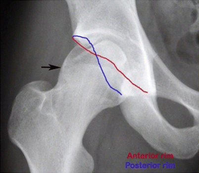
Fig. 9.3
AP radiograph of a hip
Figure 9.3 shows acetabular crossover sign at intersection of the anterior rim (red line) and posterior rim (blue line) and ischial spine sign. The ischial spine is visible due to retroversion of the acetabulum. The overall radiographic impression is of focal pincer FAI from a retroverted acetabulum. In addition a pistol-grip deformity (arrow) confirms cam-type FAI morphology. There is a further finding of a superolateral femoral head defect present in this radiograph.
Figure 9.4 demonstrates measurement of the alpha angle. On CT or MRI axial images a line is drawn from the centre of the femoral head through the middle of the femoral neck and a second line is drawn through a point where the contour of the femoral head-neck junction exceeds the radius of the femoral head. The angle between the lines is calculated. The image on the left shows a normal hip with an angle of under 55°. The image on the right shows a hip with FAI and an angle of 64°.
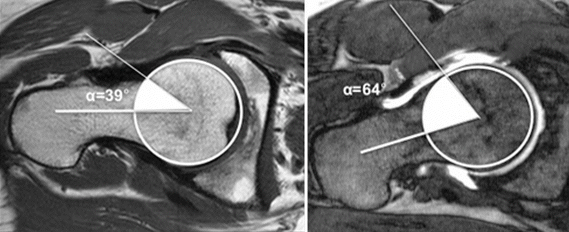
Fig. 9.4
Axial MRI of normal (left) and FAI hip (right) to demonstrate measurement of the alpha angle
CT and in particular high resolution CT with 3D reconstruction can give a clear picture of the bony anatomy as shown in figure below where a cam lesion is evident. However, MR imaging is preferred because of the lower radiation dose (Fig. 9.5).
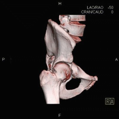
Fig. 9.5
3D CT reconstructions demonstrating a cam lesion
Plain MRI is a useful investigation to assess the bony and soft tissue anatomy. It has the advantage of avoiding radiation exposure and is non-invasive. Soft tissue inflammation from impingement, tendonitis and muscle hernias will be evident on MRI. Alternatively, an MRI arthrogram can give more detail, assesses for associated problems and differential diagnoses such damage to articular cartilage and labral tears and is considered to be a more useful investigation.
How Should FAI Be Managed?
Non-operative management can be appropriate for those with mild symptoms or activity specific symptoms. This includes physiotherapy, activity modification and analgesia. Physiotherapy should focus on strengthening the hip rather than range of movement and activities that involve stretching such as yoga should be avoided.
Intra-articular steroid and local anaesthetic injection can offer temporary relief from pain.
Surgical management options can be divided into:
Arthroscopic
Mini-open surgery
Open surgery
With improving techniques and equipment, arthroscopy offers increasingly greater access to various pathologies within the hip (Fig. 9.6). This includes:
Anterior cam lesionsRelated posts:
 Evidence-Based Treatment of Flexible Flat Foot in Children
Evidence-Based Treatment of Glenohumeral Dysplasia Caused by Obstetric Brachial Plexus Injuries
Evidence-Based Treatment of Flexible Flat Foot in Children
Evidence-Based Treatment of Glenohumeral Dysplasia Caused by Obstetric Brachial Plexus Injuries
 Evidence-Based Treatment of Deformity in Multiple Osteochondromatosis
 Evidence-Based Treatment for Slipped Upper Femoral Epiphysis
 What Is the Best Treatment for Blount’s Disease?
 Evidence-Based Treatment for Musculoskeletal Disorders in Children with Down’s Syndrome

Stay updated, free articles. Join our Telegram channel

Full access? Get Clinical Tree


