Fig. 21.1
Pes cavus foot, cavovarus type
In children, Pes Cavus is usually neuromuscular in aetiology, and half of this group has Charcot-Marie-Tooth (CMT) disease [2]; other neurologic or spinal cord conditions present similarly. These disorders give rise to muscle imbalance that produces the observed deformity. The clinical deformity in the cavovarus foot has a characteristic pattern:
weak intrinsic muscles and relatively spared extrinsics > Claw toes
weak anterior compartment (Tibialis anterior in particular) and strong Peroneus longus > plantar-flexed first ray > MTPJ extension contracture > contracted plantar fascia > cavus
weak Tibialis anterior and Peroneus brevis and unopposed Tibialis posterior > varus hindfoot
The diagnosis is clinical but there are established radiological criteria on weight-bearing lateral views of the ankle and foot. The two most commonly used measurements are the calcaneal pitch (angle between the inferior surface of the calcaneum and the floor; greater than 30° in Pes cavus) and the Meary angle (angle between the long axes of the first metatarsal and the talus; more than 5° in Pes cavus).
Questions
From a clinical perspective, we wished to establish the evidence base for the following questions (the first concerning the role of conservative management, and the latter four on modes of surgical management):
- 1.
Is there a role for conservative treatment in Pes cavus?
- 2.
What is the evidence for the treatment options in management of flexible Pes cavus?
- 3.
What are the results of joint-sparing surgery for management of rigid Pes cavus?
- 4.
What are the results of triple arthrodesis in management of rigid Pes cavus?
- 5.
What are the results of external fixation in management of Pes cavus?
Searching for Evidence
We searched MEDLINE and the Cochrane database for relevant studies and limited our search to the English language. The complete search yielded 325 references. Twelve references were deemed relevant and included for review after screening of the title and abstract. We also checked the reference list of the included articles and used the snowball method for reference harvesting. This generated nine further references for inclusion.
Search was conducted using the following strategy: (09/01/2016)
Cochrane Database with search term “(Pes Cavus OR Cavo-varus)” three citations
PubMed search: (“Foot Deformities/surgery”[Mesh]) AND (“Foot Deformities/therapy”[Mesh]) AND ((Randomized Controlled Trial[ptyp] OR Clinical Trial[ptyp] OR Comparative Study[ptyp] OR Observational Study[ptyp]) AND English[lang] AND (infant[MeSH] OR child[MeSH] OR adolescent[MeSH])) 251 citations
PubMed (www.ncbi.nlm.nih.gov/pubmed/) clinical queries search/systematic reviews: Therapy/Narrow[filter] AND ((“foot deformities”[MeSH Terms] OR (“foot”[All Fields] AND “deformities”[All Fields]) OR “foot deformities”[All Fields] OR (“pes”[All Fields] AND “cavus”[All Fields]) OR “pes cavus”[All Fields]) AND (“therapy”[Subheading] OR “therapy”[All Fields] OR “treatment”[All Fields] OR “therapeutics”[MeSH Terms] OR “therapeutics”[All Fields])) AND ((Randomized Controlled Trial[ptyp] OR Clinical Trial[ptyp] OR Comparative Study[ptyp] OR Observational Study[ptyp]) AND English[lang] AND (“infant”[MeSH Terms] OR “child”[MeSH Terms] OR “adolescent”[MeSH Terms])) 71 citations
Is There a Role for Conservative Treatment in Pes Cavus?
There is good quality evidence in support of conservative treatment (rather than no treatment) of Pes cavus, limited by its utility especially in a progressive deformity. In a randomised controlled trial, Burns et al. compared the effect of custom-made orthoses vs sham orthoses in 154 adults with symptomatic bilateral Pes cavus [3]. This trial was conducted on adults, so there are questions over its applicability to children. Foot pain was the primary outcome measure; at 3 months, participants using the custom-made orthoses reported significant reduction in foot pain (difference, 8.3 points; 95 % confidence interval [CI], 1.2–15.3 points; P = .022). Plantar pressure and functional scores were also improved (although quality of life did not).
A subsequent Cochrane review suggested that off-the-shelf orthotics did not improve clinical outcomes [4]. The same group subsequently conducted a controlled trial to investigate the use of Botulinum toxin type A (BoNT-A) in preventing the progression of cavus deformity in ten children (20 feet) with CMT type 1A [5]. The control leg received no injections. After 2 years, there was no significant difference in radiological alignment, foot posture, ankle flexibility or strength between the sides. The authors concluded that BoNT-A did not affect the progression of cavus deformity in CMT 1A.
Surgical Treatment of Pes Cavus
Surgical treatment can be divided into soft tissue (contracture release, tendon lengthening/shortening/transfer), osteotomy and arthrodesis. These are not mutually exclusive but need to be combined and tailored to the needs of the individual patient. The Coleman block test [6] is an important pre-operative decision-making tool, determining whether the hindfoot varus is correctible (flexible) or fixed (rigid). Hindfoot surgery is unnecessary in a flexible, forefoot-driven Pes cavus, but essential in a rigid hindfoot varus.
A variety of procedures have been described for treatment of both flexible and rigid deformities; no single combination has gained unanimous approval. This is understandable as surgical decision-making takes into account a multiplicity of factors, including: apex of deformity, rigidity of deformity, type of cavus, hindfoot position and flexibility, static/dynamic deformity, muscle strength and joint degeneration etc. Despite the claims of various proponents, it remains unclear as to when is the best time for surgery, or indeed whether early surgery prevents later joint degeneration (Figs. 21.2 and 21.3).
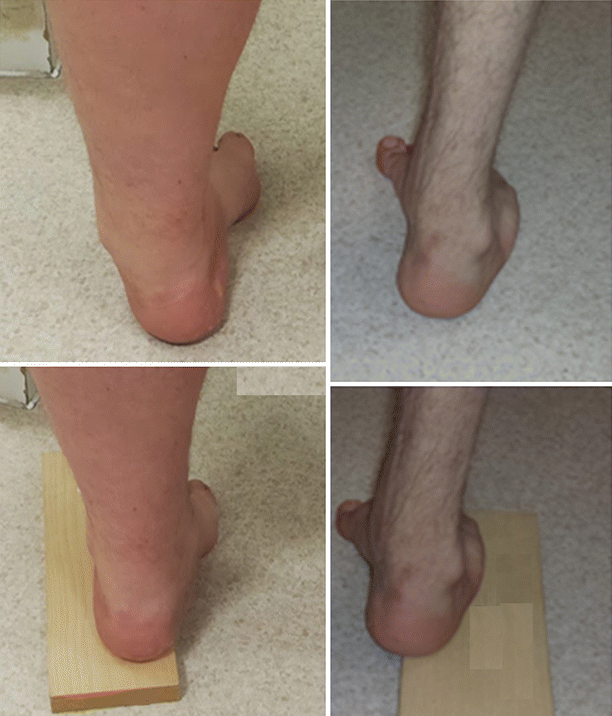
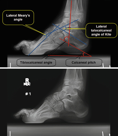
Fig. 21.2
Coleman’s test; left: hindfoot is flexible. Right: hindfoot is fixed
Fig. 21.3
Pes cavus: radiological assessment
What Is the Evidence for the Treatment Options for Management of Flexible Pes Cavus?
In flexible Pes cavus deformity, joint-sparing surgery (purely of soft tissues or with bony surgery also) can be performed, with the intent of preserving joint function. The primary outcome of interest is a pain-free mobile plantigrade foot and, in the longer term, preservation of joint function without degenerative change.
Chan et al. performed a useful study in this regard [7], investigating foot pressure distributions before and after surgical correction in nine children with CMT who had joint sparing surgery. Although surgery improved all radiological parameters, the pressure distributions remained abnormal. They concluded that pressure distribution normalisation depends on achieving a balanced foot and a correct heel position.
Roper and Tiberwal [8] reviewed the results of soft tissue surgery at a mean follow up of 14 years in ten CMT type I patients (mean age at surgery 14 years, but age range 5–36 years) who underwent open Tendoachilles lengthening (TAL), split transfer of Tibialis anterior tendon (TAT), plantar fasciotomy (PF), claw toe release with flexor-extensor transfer and modified Robert Jones procedure for claw hallux correction. Jones originally proposed Extensor Hallucis Longus (EHL) tendon transfer to the first metatarsal neck for claw toe correction [9]. Subsequent modification involved the fusion of the hallux IP joint [10]. Unfortunately, the authors did not indicate the severity of Pes cavus, nor the flexibility of the hindfoot varus; in fact, one patient underwent calcaneal osteotomy for “very severe varus”. Outcomes of interest were subjective, including function, appearance and symptoms. Global outcomes were classified as excellent, good, fair or poor. There were no complications. Two patients had recurrent deformity that required repeat soft tissue surgery. All patients had satisfactory results and were able to walk “unlimited distances” on latest review.
In an informative level IV study, Ward et al. [11] described the results (at a mean follow up of 26 years) of joint-sparing surgery in 25 patients with CMT who had a flexible hindfoot [mean age at surgery 15.5 years (8.7–25.1)]. The authors developed an algorithmic approach to flexible Pes cavus management. All patients underwent PF (to reduce the cavus deformity) and transfer of Peroneus longus (PL) tendon to Peroneus brevis (PB) (to remove the deforming force on the first ray). Most patients also underwent first metatarsal osteotomy (DFO), if the foot was deemed not plantigrade following the initial procedures. If there was clawing of the great toe patients underwent Extensor Hallucis Longus (EHL) recession. Those with pre-operative power of at least grade IV in Tibialis anterior underwent TAT transfer to the lateral cuneiform to supplement eversion strength; the transfer of the TAT was not part of the authors’ initial practice but was subsequently included, and three patients underwent secondary tendon transfer. Overall, effectively deviating from the latterly advised protocol, TAT transfer was performed to the cuboid or the middle cuneiform in nine patients (14 feet). Some patients had other additional surgery (hallux IPJ fusion – 6 feet, TAL – 1 foot). Seventeen patients (29 feet) had both clinical and radiological assessment. Seven patients (8 feet) required 11 subsequent operations of which there was one calcaneo-cuboid fusion and one ankle fusion. Eleven patients required orthosis at follow-up. General health (SF-36) score means were 49.8 (mental component score) and 37.7 (physical functioning score). Foot function was assessed using the Foot function index (FFI), having three sub-scales of pain, disability and activity limitation (maximum score is 100 with higher scores indicating worse function). Mean scores in the three sub-scales were 35, 40.5 and 22.1, respectively. Twenty-one patients had some degree of hindfoot varus although cavus correction was well maintained. Osteoarthritis (OA) was seen most commonly at the medial cuneiform-first metatarsal joint; 11 joints in 8 feet demonstrated OA.
Chatterje and Sahu reported the results of midfoot osteotomy in 18 adolescents [mean operative age 11.3 years (range: 8.6–15 years); mean follow-up 5.4 years, with no loss to follow-up] who had unilateral Pes cavus (all but one following poliomyelitis; the other having meningocoele) [12]. Patients were treated with the Japas osteotomy (midfoot osteotomy with the apex placed over the navicular) after initial open PF release. Thirteen patients required additional TAL; two patients had a rigid hindfoot. No radiological parameters were presented. Outcome was subjective, being graded as “very good”, “good” or “poor” based on completeness of deformity correction. Four patients had poor results necessitating further surgery: two underwent triple fusion and two underwent calcaneal osteotomy.
Leeuwesteijn et al. reported a series included 33 patients with CMT [mean operative age was 28 years (range: 13–59 years); only five patients were adolescents (<16 years); mean follow-up was only 57 months], with flexible hindfoot deformity [13]. All patients underwent DFO of the first metatarsal; additional surgery consisted of hallux IPJ arthrodesis (34 feet), percutaneous TAL (28 feet), claw toe correction (28 feet), Peroneus longus to Peroneus brevis transfer (27 feet), EHL transfer (15 feet), Tibialis posterior tendon transfer due to drop foot (six cases), PF release (1 foot). The authors chose to transfer EHL to the Tibialis anterior or Peroneus tertius tendons (rather than the neck of the first metatarsal), thinking that this transfer regime resulted in a lower incidence of hallux elevatus. There were no major complications. Outcomes were assessed using the FFI and patient satisfaction score: there was a statistically significant improvement in pain (from 29.3 to 14.8) and disability (from 37.8 to 23.5) components of the FFI. Ninety percent of respondents were satisfied with the deformity correction but even over this time frame, two patients underwent triple arthrodesis (TA) due to deformity recurrence.
The most recent evidence Faldini et al. [14], presenting the results [mean follow up 6 years (range: 2–13 years)] of 12 CMT 1A patients (24 feet) with bilateral foot deformities treated by PF release, midtarsal osteotomy (MTO), naviculo-cuneiform arthrodesis (NCA), Jones procedure and DFO of the first metatarsal. It is interesting that the authors do not appear to have attempted tendon transfer to balance power. They maintain that elevation of the first metatarsal head would indirectly correct the varus heel in a flexible deformity. It is notable that their pre-operative investigations reveal that 17 and 16 feet (of the 24) had 5/5 power in Peroneus brevis and Tibialis anterior, respectively; this may be why their regime was successful, in spite of the absence of tendon transfers. Five patients required additional surgery for claw toe correction. Outcomes were assessed using the Maryland foot score (MFS), rated as excellent (100–90 points), good (89–75 points), fair (74–50 points), or poor (<50 points). Mean score improved from 72 to 86 and 12 feet reported excellent results. Two feet had superficial wound dehiscence. There was no recurrence or subsequent surgery.
In summary, a sequence of level IV case series suggests joint-sparing surgery to be a viable treatment option in flexible Pes cavus. There are no universally agreed guidelines – patients need careful assessment and treatment options should be individualised. Current evidence appears to suggest that a combination of soft tissue and bony procedures are necessary. In flexible Pes cavus, there is inadequate evidence to determine if joint sparing procedures delay progression of deformity or subsequent joint degeneration. Patients should be counselled that further treatment may be necessary (Table 21.1).
Table 21.1
Summary of the results of surgery for management of flexible Pes cavus (all level IV case series)
Study | N (feet) | Diagnosis | Treatment | Follow up in years Mean (range) | Re-operations (triple fusion) | Outcome |
|---|---|---|---|---|---|---|
Roper and Tiberwal [8] | 10 (18) | CMT Type 1 | TAL, split transfer of TAT, PF, claw toe release and modified Robert Jones procedure | 14 (6–32) | 3 (0) | All “satisfactory” |
Ward et al. [11] | 25 (41) | CMT | PF, Transfer to PL to PB, EHL recession, transfer of TAT, DFO 1st MT | 26 (9.9–33.5) | 11(0) | 21 hindfoot varus Significant OA in 8 feet (11 joints) |
Chatterjee and Sahu [12] | 18 (18) | Post Polio 17 Meningocele 1 | Japas osteotomy (included PF) Tendo Achilles lengthening [13] | 5.4 (3–13) | 4 (2) | Very good: 6 Good: 8 Poor: 4 |
Leeuwesteijn et al. [13] | 33 (52) | CMT | DFO 1st MT and soft tissue surgery | 4.7 (1–13) | 10(2) | 90 % satisfied with deformity correction |
Faldini et al. [14] | 12 (24) | CMT 1A | PF, MTO and NCA Jones procedure and DFO 1st MT | 6 (2–13) | 0 | Mean MFS improvement: 14 points |
What Are the Results of Joint Sparing Surgery for Management of Rigid Pes Cavus?
For a rigid deformity, earlier procedures attempted only uniplanar or biplanar correction. More recently, techniques have focussed on achieving multiplanar correction. Most of these operations involve a midfoot osteotomy that heals – either by arthrodesis, pseudoarthrodesis or bony union. Technically, these may not be joint-sparing surgery, but most preserve the Chopart joint complex.
Related posts:
 Evidence-Based Treatment of Flexible Flat Foot in Children
Evidence-Based Treatment of Glenohumeral Dysplasia Caused by Obstetric Brachial Plexus Injuries
Evidence-Based Treatment of Flexible Flat Foot in Children
Evidence-Based Treatment of Glenohumeral Dysplasia Caused by Obstetric Brachial Plexus Injuries
 Evidence-Based Treatment of Deformity in Multiple Osteochondromatosis
 Evidence-Based Treatment for Slipped Upper Femoral Epiphysis
 Private: Physeal Injury, Epiphysiodesis and Guided Growth
 Femoro-Acetabular Impingement in Children
Stay updated, free articles. Join our Telegram channel

Full access? Get Clinical Tree


