Fig. 40.1
Equinus feet in a child with cerebral palsy
Does Botulinum Toxin Injection Correct Spastic Equinus Deformity and Improve Gait or Function?
Botulinum toxin is produced by the anaerobic bacterium Clostridium botulinum and selectively blocks the release of acetylcholine at the cholinergic nerve terminal of a neuromuscular junction. This causes a temporary reduction in muscular activity in the specific muscles [6]. Botulinum toxin intramuscular injection has been used to reduce the spasticity of specific muscle groups in children with cerebral palsy. The reduction of spasticity would augment the effect of interventions (physiotherapy, serial casting, orthotics) that prevent the formation of contractures, enhance motor ability and improve function [7].
Botulinum toxin has been used extensively to address the spastic equinus foot deformity. The types of botulinum toxin, dosage, number of sites of injection and use of adjunct interventions (casting, physiotherapy) vary significantly in the literature. Current evidence supports the use of botulinum toxin to correct the spastic equinus deformity in ambulatory children with cerebral palsy. It has been shown that it improves gait and may improve function.
In a randomized double-blind control study, Koman et al. evaluated the effect of botulinum toxin in 12 ambulatory children with cerebral palsy and dynamic equinus contracture [8]. They assessed gait and function using observational gait analysis, a physician rating scale and a parent/guardian questionnaire. The children that were treated with botulinum toxin had significant improvement in their gait pattern compared to the placebo group.
In a multi-center randomized double-blind control study, Koman et al. evaluated the effect of botulinum toxin in 114 ambulatory children with cerebral palsy and dynamic equinus using an observational gait analysis scale (Physician Rating Scale) [9]. The patients were evaluated in five occasions up to 12 weeks post treatment. Again, the group of patients treated with botulinum toxin demonstrated improved gait pattern comparing to the placebo group. The positive effect on gait pattern lasted up to 12 weeks post treatment.
Sutherland et al. used instrumented gait analysis to objectively evaluate the gait of children with dynamic equinus [10]. In this prospective, randomized, double blind clinical trial, 20 children were evaluated before and 8 weeks following treatment. The children that were treated with botulinum toxin showed improved gait kinematics (ankle dorsiflexion in stance and swing) in the short term.
Twelve matched pairs of children with spastic hemiplegia and dynamic equinus contracture were used in a pair randomized controlled clinical trial to measure the effect of botulinum toxin in functional ability [11]. Love et al. measured motor function using the Gross Motor Function Measure (GMFM) on enrolment and at 1, 3 and 6 months post injection. The children that had treatment with botulinum toxin demonstrated reduced spasticity and improved motor function that lasted up to 6 months.
Bjornson et al. reported similar findings [12]. In a randomized double masked placebo controlled study, they measured the gross motor skills (GMFM) and energy expenditure in 33 ambulatory children with spastic diplegia without fixed equinus contractures. Seventeen participants were randomized to receive bilateral gastrocnemius botulinum toxin injections and 16 received saline injections. The group of children that received botulinum toxin demonstrated improved gross motor skills 6 months following treatment.
The effect of botulinum toxin on walking was assessed with a video gait analysis scale by Ubhi et al. [13]. In a randomised double blind placebo controlled trial, the gait and function of forty ambulatory children with cerebral palsy and spastic equinus deformity was assessed in four occasions (baseline, 2,4 and 12 weeks post treatment). The children that were treated with botulinum toxin demonstrated improved gait pattern at 6 and 12 weeks; improved walking function (GMFM) at 12 weeks post treatment.
In another randomized control study, El-Etribi et al. compared 20 children with spastic diplegia and dynamic equinus that were treated with botulinum toxin injections and physiotherapy with a similar group of children that were treated with physiotherapy alone [14]. The authors assessed spasticity, ankle range of motion and gait pattern using an observational gait analysis scale (Physician Rating Scale) pre and post treatment. The follow-up of these patients was 12 weeks after treatment. The children that were treated with botulinum toxin injections had reduced spasticity and improved gait pattern that lasted at least for 12 weeks.
Different modalities have been used as an adjunct to botulinum toxin injections, in order to enhance its beneficial effect. The use of serial casting, orthotics and physiotherapy has been suggested to augment the effects of botulinum toxin injections.
Two randomized control studies evaluated the adjunctive effect of casting following botulinum toxin injection in children with dynamic equinus. Bottos et al. compared five children with spastic diplegia that were treated for dynamic equinus with botulinum toxin injections alone with five that had casting for 3 weeks post injection [15]. The addition of casting provided longer lasting effect of reduced spasticity and improved function (GMFM) at 4 months post treatment and increased stride length.
In a multicenter randomized study, 39 ambulatory children with cerebral palsy and dynamic equinus were divided into three treatment groups [16]. One group was treated with botulinum toxin injections alone, the second with botulinum toxin injections and casting and the third with placebo injections and casting. The two groups of patient that were treated with casting showed improvements in ankle kinematics, spasticity, passive ankle range of motion and dorsiflexion strength. No significant differences were identified on the patients that were treated with botulinum toxin injections alone.
Physiotherapy programs are commonly used in children with cerebral palsy. These though differ a lot in terms of the modalities used and their intensity. There is good evidence that physiotherapy programs such as strengthening programs are effective in children with cerebral palsy [17]. Physiotherapy has been part of all studies that evaluated the effectiveness of botulinum toxin injections. There is insufficient evidence that can support or refute the use of physiotherapy programs as an adjunct to botulinum toxin injections to improve dynamic equinus in children with cerebral palsy. Most experts would suggest that physiotherapy should be always be used when treating dynamic equinus deformities in children with cerebral palsy using botulinum toxin injections. There is consensus among experts that physiotherapy modalities, such as stretching, strengthening, targeted motor training should be used routinely as adjuncts to treat dynamic equinus with botulinum toxin injections [18, 19].
Does Surgical Treatment of Equinus Deformity Improve Gait and Function in Ambulatory Children with Cerebral Palsy?
Dynamic equinus deformity in children with cerebral palsy progresses with time into a static contracture of the gastrosoleus complex. Equinus contracture is usually part of a constellation of lower limb deformities that affects the gait of children with cerebral palsy. Equinus contracture can impair gait efficiency, lead to foot pain and difficulties with orthotic use and footwear. When non-operative measures fail to control and correct an equinus contracture, surgical treatment is indicated.
Various surgical techniques to lengthen the gastrosoleus complex have been described. Their main difference is the level of lengthening of the gastrosoleus complex: it can be at the muscle belly of gastrocnemius, at the gastrocnemius aponeurosis and soleus fascia or at the Achilles tendon. Lengthening at the level of gastrocnemius muscle belly is indicated when only this muscle is involved. When both the gastrocnemius and soleus are affected, more distal lengthening is performed.
With procedures that are more distal, more lengthening can be achieved, but there is higher risk of over-lengthening and muscle weakness. The outcome of surgical correction of an equinus contracture is more predictable as a child gets older [23]. Children with hemiplegia are more prone to recurrence of the equinus contracture, while children with diplegia are more prone to over-lengthening and developing a calcaneus deformity [24, 25].
Surgical correction of equinus contracture in children with cerebral palsy improves gait and function in the short and long term. It is usually part of a multi level surgical intervention (single event multilevel surgery) to address all possible dysfunctions that affect gait and function. Davids et al. evaluated the results of surgical lengthening of the gastrocnemius-soleus complex of 53 children with cerebral palsy in a retrospective, cohort study [26]. The mean time between the initial and postoperative follow-up study was 2 years and 3 months, while the follow-up assessment was performed between 1 and 3 years following surgery for most of the children. Significant improvements in swing phase kinematics (mean and peak ankle dorsiflexion) were recorded with three-dimensional gait analysis following surgery.
Saraph et al. evaluated the outcome of 22 children with spastic diplegia that had Baumann gastrosoleus recession, as part of a single event multilevel surgery [27]. The function of the ankle showed significant improvement, as this was assessed by clinical examination and gait analysis before and at 2 years after the operation.
Forty children with spastic diplegia were followed-up for a mean of 7.5 years after gastrocnemius recession or differential gastrocnemius-soleus complex lengthening, as part of single-event multilevel surgery [28]. There was statistically significant and clinically relevant improvement in ankle kinematics and kinetics following surgery. There was a significant improvement in gait function, as this is measured with the Gait Profile Score. The authors reported a rate of recurrence of 35 % and overcorrection of 2.5 %.
In a similar study, Dreher et al. reported on the outcomes of 44 children that had gastrocnemius-soleus recession surgery as part of a single event multi-level surgery [29]. The mean follow up assessment of these children was 8.6 years following the index procedure. The authors reported 24 % recurrence rate and 11 % overcorrection. Kinematic and kinetic data showed significant improvements that were maintained long-term. The maximum ankle power in stance phase was reduced 1 year after surgery, but returned at 3 years follow-up and remained at the 9 years follow-up. Most of the gait parameters showed a tendency for deterioration between the 1-year and 9-year assessments, although these differences did not reach statistical significance.
Svehlik et al. also reported this tendency for deterioration with long-term follow-up [30]. Eighteen children with spastic diplegia that underwent Baumann procedure to correct equinus gait were evaluated clinically and with instrumented gait analysis 10 years following their surgery. There was a significant improvement of ankle kinematics and kinetics following surgery. Despite the tendency for deterioration with time, these remained significantly improved.
Shore et al. performed a systematic review of the literature [31]. They found very poor levels of evidence. Most studies were level 4 quality of evidence, leading to only grade C recommendation. The true of recurrence and overcorrection rate is difficult to detect, since literature includes heterogeneous groups of patients with short-term follow-up. The literature indicates greater incidence of recurrent equinus in children with hemiplegia regardless of procedure, and greater incidence of overcorrection in children with diplegia, particularly following procedures on the Achilles tendon.
There are few studies comparing the different surgical techniques used to correct an equinus contracture in children with cerebral palsy. Most of them are retrospective studies, not randomized, with short follow-up and non-standardized selection criteria. The choice of surgical technique depends on the involvement of the gastrocnemius only or the whole gastrosoleus complex in the equinus deformity. Decision can be made using the Silfverskiöld’s test and the instrumented gait analysis data. In children with hemiplegia, there is usually contracture of both the gastrocnemius and soleus. Percutaneous and open Achilles tendon lengthening has been shown to have equally satisfactory outcomes [25]. More distal procedures should be used in children with spastic diplegia, where only the gastrocnemius is usually contracted and the natural history is a progression to a calcaneus deformity [32]. They have a higher risk of recurrence but a low risk of overcorrection [29]. It is always easier to correct a recurrent equinus contracture than to manage a calcaneus deformity.
Equino-Cavo-Varus Deformity
Equinovarus and equinocavovarus deformities are common foot deformities in children with cerebral palsy (Fig. 40.2). The deformity consists of heel varus and equinus with midfoot adduction and supination. Cavus may be also part of the deformity, as the forefoot is plantar-flexed in relation to the hindfoot varus. These deformities are believed to be the result of the muscle overactivity of the tibialis posterior, tibialis anterior or both. This deformity is common in younger children with cerebral palsy. It usually progresses to a fixed deformity with time in children with spastic hemiplegia, while in diplegic or quadriplegic children it usually overcorrects into valgus [33].
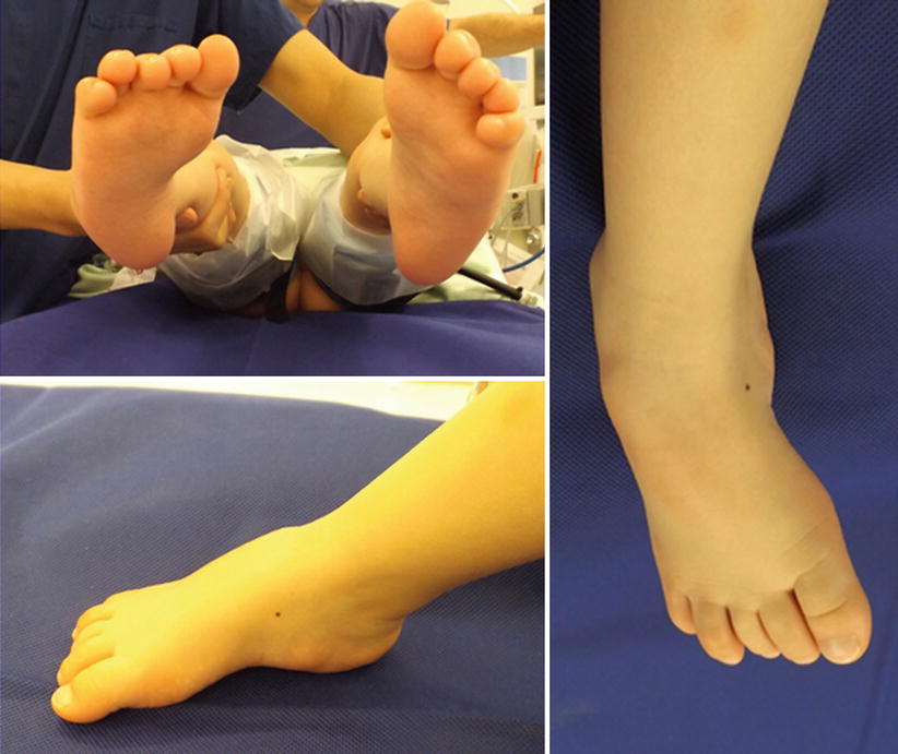
Fig. 40.2
Equino-cavo-varus deformity
This deformity can cause difficulties with shoe wear, brace intolerance and affect walking. As the deformity progresses and become more rigid, children stand on the lateral border of the foot. This results into painful callus formation or even fifth metatarsal stress fractures.
Orthotics can be used in flexible deformities in young children, since these are usually stable in hemiplegic children and may overcorrect into valgus in diplegic or quadriplegic patients [33]. In older children, with flexible or rigid deformities that affect gait, cause pain or interfere with bracing, operative treatment is usually indicated.
Does Surgical Treatment of Equinovarus Deformity Improve Gait in Ambulatory Children with Cerebral Palsy? Which Is the Best Surgical Technique?
The evidence for operative treatment of equinovarus foot deformities in children with cerebral palsy in unfortunately poor. It consists mainly of case series with short follow-up (Level 4) and review articles from experts (Level 5). There are no randomized or cohort comparative studies.
Flexible deformities are usually addressed with soft tissue surgery (soft tissue release, tendon lengthening or tendon transfer), while rigid deformities require bony surgery (osteotomies or joint fusion).
Flexible deformities can be corrected with tibialis posterior lengthening, split tibialis posterior transfer, split tibialis anterior transfer or combination of these. Tendon transfer of the whole tendon should be avoided, as there is a high risk of overcorrection, especially in diplegic and quadriplegic patients [34–36]. Lengthening of the tibialis posterior can be performed either proximally as an intramuscular lengthening or distally as Z-lengthening of the tendon [37, 38].
Split transfer of the lateral part of the tibialis anterior or posterior tendon into the lateral part of the foot is preferred [39, 40]. There is lower risk of overcorrection and the muscle does not lose its power. The choice depends on which muscle is the main contributor of the deformity. Michlitsch et al. reviewed the gait analysis data of 78 patients with cerebral palsy and varus foot deformity [41]. The varus foot deformity was a result of the dysfunction of the tibialis anterior in 34 %, tibialis posterior in 33 % and both in 31 %. In the majority of the cases varus occurred during both stance and swing phase of the gait. The timing of varus during the gait is a poor predictor of the associated muscle dysfunction. The use of dynamic electromyography and kinematic data can assist to determine the main contributing muscle to the foot deformity [41, 42].
Most of the studies demonstrate satisfactory outcomes in the short and medium term [40, 43–46]. Studies with longer follow-up show that the outcomes seem to deteriorate with time (recurrence or overcorrection). Kling et al. reported the outcomes of 37 split tibialis posterior transfer in 31 patients with spastic cerebral palsy [40]. The mean follow up of these patients was 8 years. They reported three poor results.
Eighteen children that had split tibialis posterior transfer through the interosseous membrane [47]. The mean follow up was 8.4 years. The authors reported just one poor result.
Chang et al. have reported the largest series of children with spastic equinovarus foot deformities [38]. One hundred and eight children (140 feet) with a mean follow up of 7.3 years were assessed. The surgical techniques used were split tibialis posterior tendon transfer or Z-plasty of the tendon at the level of the medial malleolus or intramuscular lengthening. Sixty-five patients were considered as failures of treatment (recurrence or overcorrection). The factors that had an effect on surgical outcome were involvement of cerebral palsy, age at operation and preoperative ambulation status. Hemiplegic patients, children older than 8 years at the time of operation and ambulatory status have more favorable outcome comparing to diplegic/quadriplegic involvement, younger children and non-ambulators. None of the surgical techniques used was found to give better outcomes.
Flexible equinovarus deformities can progress into rigid deformities with time. Fixed deformities require bony surgery; this can be in the form of osteotomies for mild deformities or arthrodesis for severe ones. Fixed hindfoot deformities can be addressed with calcaneal sliding or closing wedge osteotomy. Cavus and rigid supination deformities can be corrected with midfoot (medial cuneiform, cuboid) osteotomies or joint fusions.
For severe rigid deformities, a triple fusion provides the most reliable long-term outcomes. Tenuta et al. reported on ten patients with cerebral palsy that had triple fusion for a rigid equinovarus foot deformity [48]. The mean follow-up was 16.1 years. Nine patients had no residual deformity; three patients were dissatisfied with the outcome, six reported foot pain and five had ankle degenerative arthritis findings on x-ray. In three patients, their ambulatory capacity improved, while the others remained stable.
Satisfactory outcomes following triple fusion in adult patients with cerebral palsy were also reported by Trehan et al. [49]. Seven of the 21 patients had a foot varus deformity. The mean follow-up was 22 years. Twenty patients were satisfied with the outcome, eight patients reported pain with ambulation, 10/26 feet had residual deformity and three patients had x-ray findings of ankle arthritis (only one symptomatic).
Current evidence shows satisfactory outcomes with surgical correction of equinovarus foot deformities in children with cerebral palsy in the short and mid-term. Satisfactory outcomes should be expected in older children with hemiplegia. Gait analysis studies with the use of dynamic electromyography can help to distinguish if the tibialis posterior, anterior or both are dysfunctional. The appropriate surgical technique can be used to rebalance the foot. Fixed deformities should be corrected first with appropriate osteotomies or joint fusions. In severe rigid deformities, triple arthrodesis can provide the most reliable long-term outcomes.
Equino-Valgus and Plano-Valgus Deformity
This is probably the commonest foot deformity in children with cerebral palsy and the severity can range from a very subtle collapse of medical arch to a very severe deformity where the forefoot pointing outward (Fig. 40.3). The natural history of this deformity overlaps with the natural history of flat feet in early childhood and the progress and future developments are extremely unpredictable before 8 years old [33, 50].
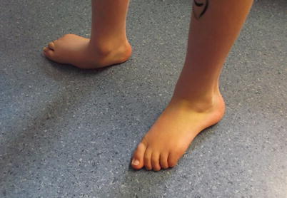
Fig. 40.3
Severe equino-plano-valgus feet deformity
Do Orthoses Improve the Outcomes of Equinovalgus Deformity in Children with Cerebral Palsy?
Tight gastrosoleus complex play an important aetiological role in the development of equinovalgus foot; hence treating this tightness may reduce the incidence and or the severity of the equinovalgus deformity. This has been covered in earlier sections and the values of orthoses have been highlighted. Moreover, the natural history of equinovalgus foot is unpredictable in early childhood (under the age of 8), therefore temporizing interventions such as orthoses are valuable while the clinical pictures are getting clearer [33, 50].
The evidence behind orthotic treatments in plano-valgus feet in normal children was discussed in Chap. 19 and concluded that they may have detrimental effects. However, in children with cerebral palsy the contrary is true. Several studies showed that ankle and foot orthoses (AFO) improve symptoms, slow deterioration and increase gait efficiency [51–54]. However, the best type of AFO has been contested. Kadhim and Miller [55] recommended a pragmatic approach to the AFO choices in children with equinovalgus deformity admitting the lack of the scientific documentation behind such treatment. They recommend a solid AFO until the child begins to walk. Afterward, a hinged (articulated) AFO is used to allow for ankle dorsiflexion and to prevent ankle plantarflexion. Two exceptions are when the child has a severe crouch posture or severe equinovalgus deformity. In the former a solid AFO (or even GRAFO in heavier children >25 kg) is more useful to improve crouch posture. In the latter, hinged AFO is less likely to provide enough support and often causes rubbing and skin irritation. They also recommended supramalleolar AFO for children who can control ankle plantarflexion and dorsiflexion.
Does Surgical Treatment of Equinovalgus Deformity Improve Gait in Ambulatory Children with Cerebral Palsy? Which Is the Best Surgical Technique?
Surgery is indicated when AFOs fail to achieve the desired purposes of keeping the feet symptoms free, providing stability for transfer and mobility. There is a natural selection in treating this deformity when ambulatory patients (GMFCS levels I, II, III) usually present early with a less severe deformity in comparison to non ambulatory patients (GMFCS level IV and V). The aim of treatment in ambulatory patients is to restore normal alignment while preserving joints mobility. Several operations and techniques have been described using a combined soft tissue and bony surgery to restore anatomy to normal or near normal. These include calcaneal lengthening (often called lateral column lengthening) (Fig 40.4), triple C osteotomy (includes medial calcaneal slide, cuboid opening wedge, cuneiform planar closing wedge), talonavicular fusion, subtalar joint fusion and triple arthrodesis. These are often combined with soft tissue procedures such as gastrocnemius muscle lengthening, peroneus brevis tendon lengthening, and tibialis posterior advancement [50, 55–61]. There is no convincing evidence that any is superior to the others. This is not unexpected, given the spectrum of the deformity and the functional demand of patients. Although some studies tried to compare different combination of surgical interventions in treating the calcaneo-valgus feet, their comparison was undermined by the fact the two comparators were not identical.
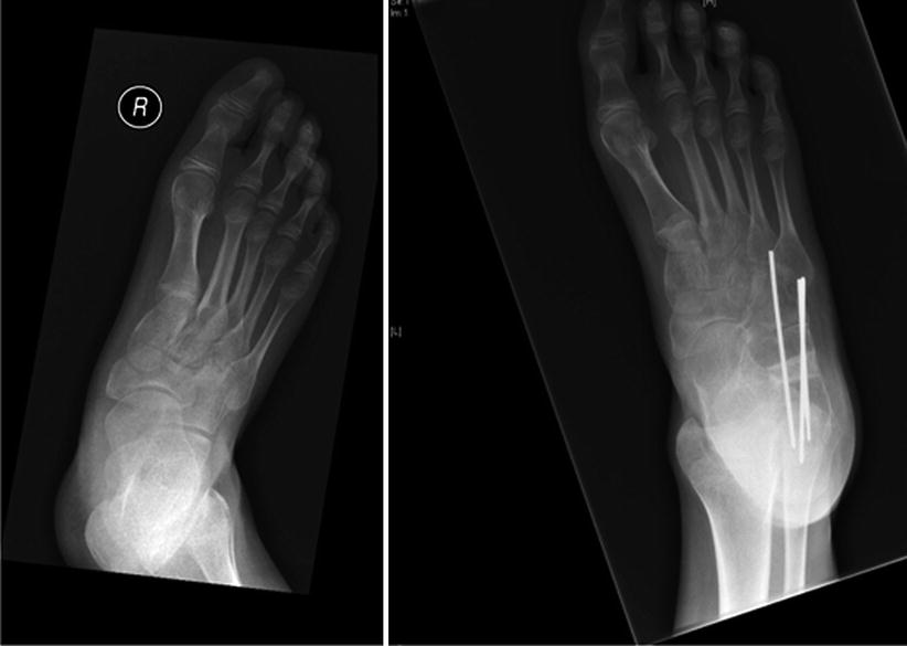
Fig. 40.4
Lateral column lengthening in a child with cerebral palsy
Mosca [62] reported on 31 patients (26 were neuromuscular) with severe, symptomatic valgus deformities of the hindfoot who were corrected with a modification of the calcaneal lengthening osteotomy. Additionally, an opening-wedge osteotomy of the medial cuneiform was used to correct the deformities of both the hindfoot and the forefoot in the patients who had a skew foot. Satisfactory clinical and radiographic correction of all components of the deformity of the hindfoot was achieved in all but the two most severely deformed feet although these two feet had sufficient correction to eliminate the symptoms.
Ettl et al. [58] reviewed the outcome of calcaneal lengthening for the treatment of planovalgus foot deformity in 19 children (28 feet) with cerebral palsy. There were 14 ambulating (19 feet) and 5 non ambulating children (9 feet). They found satisfactory results in 75 % of the feet. They found no overcorrection but a relapse of the deformity in seven cases.
Sung et al. [57] showed in a study of 71 patients with cerebral palsy that for patients with greater than 23° AP talus-first metatarsal angle, 36° lateral talus-first metatarsal angle, and 72 % naviculocuboid overlap (Fig. 40.5), additional procedures for medial stabilization, such as tibialis posterior tendon reefing and talonavicular arthrodesis, should be considered as a result of the possibility of under correction with calcaneal lengthening alone.
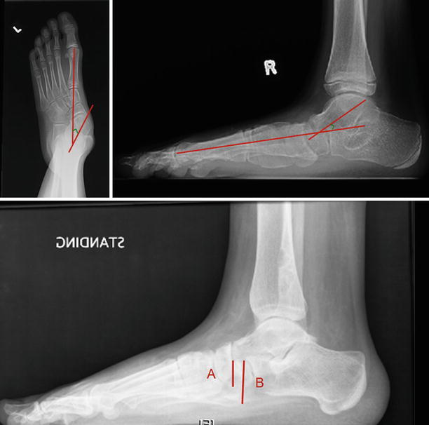
Fig. 40.5
Predictors for under correction using calcaneal lengthening alone. Top left image: AP talus-first metatarsal angle, top right image: 36° lateral talus-first metatarsal angle, and bottom image: naviculocuboid (A/B) which is the overlapped portion of the navicular and cuboid divided by the vertical height of the cuboid
Kim et al. [59] compared the clinical and radiographic results between calcaneal lengthening (18 patients, 28 feet) and triple C osteotomy (20 patients, 32 feet). The aetiology of the foot deformity was idiopathic in 16 feet and cerebral palsy in 44 feet. Non ambulatory patients were excluded. All operations were performed by a single surgeon. It was stated that the choice of surgery was random. In the calcaneal lengthening group, 19 of the 28 feet (68 %) showed a satisfactory outcome and 9 (32 %) an unsatisfactory outcome. In the triple C osteotomy group, 28 of the 32 feet (88 %) showed a satisfactory outcome and 4 (12 %) an unsatisfactory outcome. They concluded that triple C osteotomy is a more effective procedure than calcaneal lengthening for the correction of planovalgus deformity, especially severe pes planovalgus deformities.
Kadhim et al. [50] reported a long term follow up on 24 patients (43 feet); 15 were treated with calcaneal lengthening (mostly GMFCS levels I and II) and 28 with subtalar fusion (mostly GMFCS levels III and IV). Improvement was observed in both groups but additional surgery was required more among patients who were treated with subtalar fusion. Interestingly, residual foot pain was less common among children with poor functional abilities and patients who underwent subtalar fusion.
De Coulon et al. [60] questioned the reliability of other bony surgery in correcting severe planovalgus feet and they investigated the talo-navicular fusion which is used in adult practice but not in children practice. He reviewed 29 feet who were treated with talonavicular arthrodesis and reported satisfactory outcome in 28 feet, whereas 1 had unsatisfactory results according to the Yoo clinical outcome scoring scale.
Guven et al. [61] in a small series of 9 patient reported a 78 % satisfaction rates after Grice extra articular subtalar joint fusion in spite of the mean AOFAS hindfoot score increased by 15 points only from 53 (range 41–81) to 68.4 (range 51–96).
The above various case series indicate that restoring the anatomy and maintaining the correct anatomy is a key for a successful outcome. Having critically analyzed the above evidence and other excluded studies, it is impossible to favor one operation over other. Even in our hands, we do not use a single approach for all but rather tailor the treatment to individual patient and foot criteria. The deformity is multi-dimensional involving the hindfoot, midfoot and forefoot. For optimal result, a holistic approach is essential. We correct the hind foot first, then reassess the foot. There is often an associated supination deformity which requires correction. Muscles balance should then be restored to prevent recurrence. We prefer fusion surgery in severe deformity in non ambulatory children.
Foot Deformity in Children with Spina Bifida
The incidence of Spina bifida has significantly decreased in recent years (1.9/10,000 births), nonetheless it is still the cause for chronic disability in 70,000–100,000 persons in the United States with approximately 1500 new pregnancies affected per year [63, 64].
Foot deformities are extremely common in patients with Spina Bifida. Most of the children have or will develop a foot deformity whose severity is mainly related to the level of the lesion [65–67]. These deformities are particularly difficult to manage, because of the combination of motor paralysis/spasticity and sensory loss. Deformities can be present at birth in the form of Equino-Cavo-Varus (CTEV) or vertical talus, or can develop during childhood due to muscle imbalance, forces applied to the foot by weight bearing or gravity, or changes in the neurological function. Nearly all these children require some sort of treatment for their foot deformity during their lives. The goal of treatment in ambulatory patients is to provide the child with a splintable, plantigrade, supple foot able to facilitate ambulation. In non-ambulatory patient, the aim of treatment is to allow satisfactory shoe wear and positioning of the foot on the wheelchair [64, 67].
What Is the Best Treatment of Foot Deformities in Spina Bifida?
Foot deformities in Spina Bifida may present as calcaneus, equinus, varus, valgus, or a combination of deformities (Fig. 40.6). Clubfoot and vertical talus are also quite common [64–67]. Early intervention with casting, bracing, or surgical treatment may prevent fixed bony deformities. Patients with spina bifida may require several corrective procedures in order to achieve a plantigrade more functional and braceable foot [64, 68]. Roach et al. have recently reviewed 84 adults with myelomeningocele and found surgeries to maintain a plantigrade foot were helpful, because even those with low-level spinal lesions and good strength had trouble ambulating on deformed feet. However, a plantigrade foot still had a substantial risk of developing pressure sores, many progressing to deep infections and occasional amputations [69].
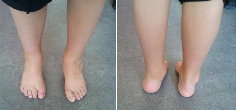
Fig. 40.6
Feet deformity in a child with spina bifida. This young boy with low level spina bifida developed right cavo-varus foot and left plano-valgus foot
Equinus Deformity
It has been described in all levels of involvement but it is mostly common in patients with no activity below the knee, i.e. thoracic and high lumbar level of involvement [65–67, 70, 71]. Unopposed gravity or the spastic activity of the triceps surae seems to be the leading causes for this type of deformity [65, 70].
Sharrard et al. suggested regular passive stretching combined with night-time splints starting at birth in an attempt to prevent the deformity in a flail foot. Authors noticed that an equinus deformity with a short tight Achilles tendon developed if stretching was abandoned. In these cases they recommended a percutaneous Achilles tenotomy to correct the deformity and the position was kept with crepe bandage rather than cast or splints. They described rapid recurrence of the deformity in some cases, often associated with a flexion deformity of the toes due to associated shortness of the flexor hallucis longus and flexor digitorum tendons. In resistant cases, they described the split-transfer of the Achilles tendon to the dorsum of the foot, the excision of the inferior tibio-fibular ligament and the excision of the talus. In their series, Authors included 64 feet with equinus deformity, which required a total of 80 procedures (77 soft-tissue operations, 1 tendon transfer and 2 bony procedures) with an overall recurrence rate of 14 % [70].
More recently, other authors reported good results after Achilles tendon lengthening, passive stretching exercises and AFOs. Achilles tendon resection and posterior release has been described for refractory cases to achieve full correction maintained by AFOs during the day and night [64, 68, 72].
The consensus is that a regime of passive stretching combined with splints should be the first line treatment for equinus deformity in spina bifida. Surgery should be considered for an unbraceable foot if skin breakdown or positioning is a problem, or to achieve a plantigrade, braceable foot in a patient with the potential for ambulation. The type of surgical procedure depends on the severity of the deformity and should be tailored on the each patient [64, 68].
Equino-Cavo-Varus Deformity
It is the most common foot deformity in spina bifida. The incidence varies with the neurologic level of involvement; in fact, it has been reported to occur in 30–50 % of patients with sacral level involvement and up to 90 % of patients with thoracic or lumbar levels of involvement [66, 67, 70, 73, 74]. Many factors may contribute to the development of clubfoot in patients with spina bifida. Sharrard noted that the most severe presentations were seen in patients with level L4 of involvement with accompanied spasticity of the triceps surae and both tibialis muscles in combination with the functional absence of the peroneal muscles [70, 71].
Relapse rates of equino-cavo-varus deformity in spina bifida are high, ranging from 22 % to 68 %, regardless of the treatment method [70, 71, 73, 75–77].
Ponseti Method
Traditionally the treatment of such a rigid and severe deformity has been extensive soft-tissue release surgery. It was common opinion that non-surgical treatment using splinting, serial casting, and stretching was unsuccessful and at considerable risk of complications including skin breakdown complicated by infection and early recurrence [70, 71]. In more recent years, the Ponseti Method consisting of manipulation and serial casting initially described for the treatment of idiopathic clubfeet [78], has been used to treat neuromuscular and syndromic clubfeet including spina bifida [75, 76, 79, 80].
Janicki et al. reported their results on the treatment of syndromic and idiopathic equino-cavo-varus feet using the Ponseti Method with a minimum follow-up of 1 year. Five patients for a total of nine feet in their cohort, had spina bifida. All nine feet were corrected using the Ponseti Method. Mean number of casts for correction was 4.2 per foot; only two of them did not require a percutaneous Achilles tenotomy. In these nine feet, there were five recurrences two of which were treated with serial casting only and three of which needed a postero-medial release. The recurrence rate in patients with myelomeningocele was 55 % compared to 13 % in idiopathic clubfeet. Major surgical release was required in 33 % of the spina bifida feet and only in 6.4 % of the non-syndromic feet (significant difference) [76].
Gerlach et al. followed prospectively 16 consecutive patients with myelomeningocele (28 clubfeet) and twenty consecutive patients with idiopathic clubfeet (35 clubfeet) managed with the Ponseti Method. The average duration of follow-up was 34 months for the myelomeningocele group and 37 months for the idiopathic group, respectively. The deformity at presentation was significantly more severe in patients with spina bifida according to the Dimeglio system [81]. The Ponseti Method was successful in all the idiopathic feet and in 27 clubfeet (96.4 %) in the myelomeningocele group. A significant difference was observed in the relapse rate of the two groups, which was 68 % in the spina bifida group and 26 % in the idiopathic group, respectively. All the recurrences were initially treated with the Ponseti Method and only four of the clubfeet in the myelomeningocele group (14 %) and one of the clubfeet in the idiopathic group (3 %) required extensive soft tissue release (no statistical difference). Complications, including blister and iatrogenic distal tibia fractures, were significantly higher in the spina bifida group [75].
Related posts:
 Evidence-Based Treatment of Flexible Flat Foot in Children
Evidence-Based Treatment of Glenohumeral Dysplasia Caused by Obstetric Brachial Plexus Injuries
Evidence-Based Treatment of Flexible Flat Foot in Children
Evidence-Based Treatment of Glenohumeral Dysplasia Caused by Obstetric Brachial Plexus Injuries
 Evidence-Based Treatment of Deformity in Multiple Osteochondromatosis
 Evidence-Based Treatment for Slipped Upper Femoral Epiphysis
 What Is the Best Treatment for Blount’s Disease?
 Evidence-Based Treatment for Musculoskeletal Disorders in Children with Down’s Syndrome
Stay updated, free articles. Join our Telegram channel

Full access? Get Clinical Tree


