Fig. 22.1
Progression of physeal closure of the distal tibia
The distal fibula epiphysis starts ossifying during the second year of life and closes 12–24 months later than the distal tibial physis. The distal tibial physis grows 4 mm a year and account for 40 % of the leg longitudinal growth.
Stability is a very important concept in the management of ankle fractures. Ligaments play a major role in ankle fracture stability. In practice most surgeons base their decisions on clinical assessment and plain x-rays to assess ankle fracture stability. Although this may be adequate in most cases where the features of instability are obvious, this is not the case in all fractures leading to some patients undergoing unnecessary surgery. In children, this is even more complicated due to the presence of the physis which poses diagnostic and therapeutic challenges to the treating surgeons.
Stress tests, CT scan and MRI scan have been used to aid decision making.
Several classifications have been proposed to help understand the nature of ankle fractures, inform treatment choices and predict future outcomes. Most of these have been based on morphological description and their predictive values for instability and better treatment choice have been questioned [2, 3]. However, to understand the published evidence better, basic knowledge of these classifications is valuable.
In adult practice, there are two common classifications in use: Weber and Lauge-Hanson classification.
Weber classified ankle fractures according to the relation of the fibular fracture to the syndesmosis into: type A (below the syndesmosis), type B (at the level the syndesmosis) and type C (above the syndesmosis). Weber classification was subsequently adopted and incorporated in the AO ankle classification which was expanded into two sub-layers as shown in Table 22.1.
Table 22.1
AO ankle fracture classification
Type A | Type B | Type C |
|---|---|---|
A1 Infrasyndesmotic lesion, isolated 1. Rupture of the lateral collateral ligament 2. Avulsion of the tip of the lateral malleolus 3. Transverse fracture of the lateral malleolus | B1 Transsyndesmotic fibular fracture, isolated 1. Simple 2 Simple, with rupture of the anterior syndesmosis 3 Multifragmentary | C1 Suprasyndesmotic lesion, diaphyseal fracture of the fibula, simple 1. With rupture of the medial collateral ligament 2. With fracture of the medial malleolus 3. With fracture of the medial malleolus and a Volkmann (= Dupuytren) |
A2 Infrasyndesmotic lesion, with fracture of the medial malleolus 1. Rupture of the lateral collateral ligament 2. Avulsion of the tip of the lateral malleolus 3. Transverse fracture of the lateral malleolus | B2 Transsyndesmotic fibular fracture, with medial lesion 1. Simple with rupture of the medial collateral ligament and rupture of the anterior syndesmosis 2. Simple with fracture of the medial malleolus and with rupture of the anterior syndesmosis 3. Multifragmentary | C2 Suprasyndesmotic lesion, diaphyseal fracture of the fibula, multifragmentary 1. With rupture of the medial collateral ligament 2. With fracture of the medial malleolus 3. With the fracture of the medial malleolus and a Volkmann (= Dupuytren) |
A3 Infrasyndesmotic lesion, with postero-medial fracture 1. Rupture of the lateral collateral ligament 2. Avulsion of the tip of the lateral malleolus 3. Transverse fracture of the lateral malleolus | B3 Transsyndesmotic fibular fracture, with medial lesion and a Volkmann (fracture of the postero-lateral rim) 1. Fibula simple, with rupture of the medial collateral ligament 2. Fibula simple, with fracture of the medial malleolus 3. Fibula multifragmentary, with fracture of the medial malleolus | C3 Suprasyndesmotic lesion, proximal fibular lesion 1. Without shortening, without Volkmann 2. With shortening, without Volkmann 3. Medial lesion and a Volkmann |
Lauge-Hansen [4] classified ankle fractures according to the position of the foot at the time of impact (supinated or pronated) and the direction of the force applied to the ankle (adduction, abduction or external rotation). A pronated foot will result in tight deltoid ligament and lax lateral ligamentous complex and vice versa for a supinated foot. Lauge-Hansen indicated that these two elements (the position of the foot and the direction of the force) determine the order in which ankle stabilising structures fail, and that these structures fail in a predictable order (Table 22.2). Several biomechanical studies failed to reproduce the work and classification of Lauge-Hansen [5, 6]). Moreover, findings from MRI studies of displaced ankle fracture did not have the patterns of ligament and bony injury predicted by their apparent Lauge-Hansen type [1, 2, 7].
Table 22.2
Lauge-Hansen classification
Lauge-Hansen class | Sequence of structures failure with increasing force caused by an injury |
|---|---|
Supination – external rotation (SER)(foot is supinated and the force is external rotation) | Stage I: ATiFL rupture or avulsion fracture of tibia or fibula |
Stage II: short oblique fibula fracture (anteroinferior to posterosuperior) | |
Stage III: PTiFL rupture or avulsion of posterior malleolus | |
Stage IV: Medial malleolus transverse fracture or disruption of deltoid ligament | |
Supination – adduction (SA) | Stage I: Talofibular sprain or distal fibular avulsion |
Stage II: Vertical medial malleolus and impaction of anteromedial distal tibia | |
Pronation – abduction (PA) | Stage I: Medial malleolus transverse fracture or disruption of deltoid ligament |
Stage II: ATiFL rupture or avulsion | |
Stage III: Transverse comminuted fracture of the fibula above the level of the syndesmosis | |
Pronation – external rotation (PER) | Stage I: Medial malleolus transverse fracture or disruption of deltoid ligament |
Stage II: ATiFL disruption | |
Stage III: Lateral short oblique or spiral fracture of fibula (anterosuperior to posteroinferior) above the level of the joint | |
Stage IV: Posterior tibiofibular ligament rupture or avulsion of posterior malleolus |
Salter and Harris introduced a classification system which carries their name based on the relationship of fracture lines to the growth plate [8, 9]. The classification is relatively simple and easily remembered and has been relatively successful in predicting future growth disturbance although the latter has been contested [10]. (Fig. 22.2 )
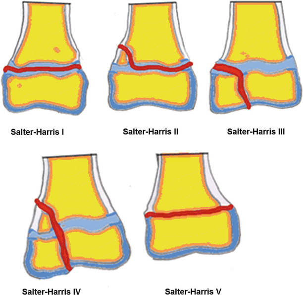
Fig. 22.2
Salter-Harris classification of physeal injury
Dias and Tachdjian [11] modified the Lauge-Hansen classification to include the Salter-Harris classification so that it can applied to children ankle fractures. Subsequently four other types of fractures were added, namely the Tillaux, triplane, axial compression, and miscellaneous physeal fractures [12].
Tillaux fracture and triplane fractures have been added to the classification. The juvenile Tillaux fractures (to differentiate it from adult similar avulsion fracture) are SH-III fractures involving the anterolateral aspect of the distal tibia plafond which is not fused to the metaphysis yet (Fig. 22.3). These fractures are caused by external rotation forces and can be reduced by internally rotating the foot [13].
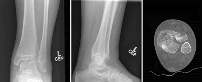
Fig. 22.3
Tillaux fracture
The triplane fracture was described by Lynn in 1972 [14]. As the name implies these fractures occur in three different planes: coronal, transverse, and sagittal. On the AP radiographs the fracture appears as a SH-III (like Tillaux fracture) whilst on the lateral view it appears as a SH-II (also called two-part triplane fracture) or SH-IV (also called three-part triplane fracture). Four-part triplane was also described in the literatures [15, 16] (see Figs. 46.1 and 46.2 in Chap. 46).
The prognostic value of these classifications has been debated. Leary [10] retrospectively reviewed 124 children after physeal fractures of the distal end of the tibia. They defined premature physeal closure (PPC) as radiographic evidence of physeal closure as compared to the uninjured side in this patient population. Fifteen fractures (12.1 %) were complicated by PPC, 67 % of the PPC observed occurred in SH-II fractures, followed by 13 % in SH-III, 13 % in SH-IV, and 7 % in triplane fractures. They did not observe any physeal arrest in the SH-I or Tillaux fractures. They were able to demonstrate statistically significant correlations between mechanism of injury and PPC and between the amount of initial fracture displacement and the rate of PPC. For each millimetre of initial displacement, there was a relative risk of 1.15 (P < 0.01).
In another study of 49 children with physeal fractures of the distal tibia or fibula or both, the Salter-Harris classification system could not significantly predict the growth pattern [17] (Fig. 22.2).
Spiegel et al. [18] followed 184 distal tibia and/or fibula, fractures for an average of 28 months after injury using the Salter-Harris classification. They differentiated three groups according to their risk for shortening of the leg, angular deformity of the bone, or incongruity of the joint. The low-risk group consisted of 89 patients, 6.7 % of whom had complications; this group included all type I and type II fibula fractures, all type I tibia fractures, type III and type IV tibia fractures with less than 2 mm of displacement, and epiphyseal avulsion injuries. The high-risk group consisted of 28 patients, 32 % of whom had complications; this group included type III and type IV tibia fractures with 2 mm or more of displacement, juvenile Tillaux fractures (Fig. 22.3 ), triplane fractures, and comminuted tibial epiphyseal fractures (type V). The unpredictable group was made up of 66 patients, 16.7 % of whom had complications; only type II tibia fractures were included. The incidence and types of complications were correlated with the type of fracture (Salter-Harris classification), the severity of displacement or comminution, and the adequacy of reduction.
de Sanctis et al. [19]) reviewed 158 ankle fractures; 132 were treated conservatively and 26 patients underwent surgical treatment. Fibular fractures of the malleolus without epiphyseal separation or dislocation (68 patients) were excluded. Of the 158 patients, 113 (70 %) were available for an average 6-year follow-up. They compared the degree of epiphyseal separation or dislocation, the Carothers-Crenshaw classification (a classification based on the mechanism of trauma) with the Salter-Harris classification (which is based on anatomical-radiographic criteria). They reported that fractures more likely to result in permanent damage to the physis are those caused by a traumatic adduction-supination mechanism that can produce SH-III, IV, and V fractures of the distal part of the tibia; they also reported that the combination of compression and adduction may cause a SH- V injury with type III and IV fractures. However, type V lesions are often diagnosed late. In 11 of their 12 poor results, 6 were caused by adduction-supination injuries and 5 were compressive injuries.
In a large retrospective study of 376 children with distal tibial physeal injury, Schurz et al. [20] reported the outcomes after various types of Salter-Harris fractures (Table 22.3).
Table 22.3
The outcomes after various types of Salter-Harris fractures
Type of fractures | Good outcome | Poor outcome | Total |
|---|---|---|---|
SH-I | 180 | 1 | 181 |
SH-II | 107 | 6 | 113 |
SH-III | 58 | 8 | 66 |
SH-IV | 15 | 1 | 16 |
Total | 360 | 16 | 376 |
Vahvanen and Aalto [21] studied 310 children treated for ankle fractures. They were classified according to the classifications of Ashhurst-Bromer-Weber, Lauge-Hansen, and Salter-Harris. They found that grouping of the fractures according to Lauge-Hansen and Ashurst-Bromer-Weber classifications suited to adults was largely unsuccessful. Epiphyseal fractures were easily classified according to Salter-Harris. They proposed that ankle fractures in children can be roughly divided into avulsion and epiphyseal fractures. Adequately reduced avulsion fractures can be expected to heal well; epiphyseal fractures, however, may cause late complications.
What Is the Evidence Behind Ankle Fractures Investigation?
Ottawa Rule
Ankle injury is common and radiological tests are not always indicated. The Ottawa ankle rules [22, 23] have been shown to be accurate in predicting the need for radiography in the acute trauma situation in adults. They can be used by medical and nursing staff in a variety of settings, and can reduce unnecessary radiography ([24, 25]; Allerston and Justham [26–28]). Several studies showed their value in detecting ankle fractures in children [29–36]. In a review by Crocco [37] of 671 fractures, the sensitivities of the Ottawa ankle rules ranged from 83 % to 100 % and specificities from 7.9 % to 50 %. X-ray reduction rates ranged from 5 % to 44 % (pooled reduction rate 25 %, 95 % CI: 23–26 %).
Plain Radiograph
The standard plain radiographic views of the injured ankle are antero-posterior (AP), the mortise and lateral views. The mortise view is a modified AP with the ankle internally rotated so that the malleoli are in the same horizontal plane and the joint space is seen evenly on both sides of the ankle. This requires 10°–20° of internal rotation. The need for three views has been questioned. Brage [38] found that ankle fractures could be classified with two radiographic views as reliably as with three views. Four different observers independently evaluated 99 sets of ankle radiographs. The examiners classified the ankle fractures by using both the Lauge-Hansen and Weber systems. The interobserver and intraobserver variations were analyzed by kappa statistics. The study demonstrated that ankle fractures can be classified with two views, lateral or mortise, with a reliability as good as that achieved with three views. The best agreement was achieved with lateral and mortise views. Adding a true AP view did not add useful information.
In a study by Vangsness [39], 123 sets of emergency room ankle x-rays (AP, lateral and mortise) were retrospectively reviewed to determine whether all three views were necessary to diagnose the presence of an ankle fracture. Four physicians (two orthopaedic surgeons, one musculoskeletal radiologist, and one emergency room physician) reviewed all randomly ordered sets of films twice – once with all three views and once with only the lateral and mortise views. The overall accuracy of two views was within the 95 % expected threshold of accuracy using three views. The lateral and mortise views alone appear sufficient for ankle fracture diagnosis, and imply a substantial decrease in radiation and cost savings.
The Role of Medial Clear Space
The presence of an ankle fracture on plain x-ray is not an indication for surgery and stable fractures may do not even require cast protection. Signs of instability are more important than the presence of a fracture as such. Displacement is often used to indicate instability (Fig. 22.4). Murphy et al. [40] measured medial and superior clear space in 73 patients without ankle injuries. Seventeen percent of male x-rays and 1 % of female x-rays had a medial clear space >4 mm, while 2 % of males and no females had a medial clear space >5 mm. Thirteen percent of radiographs had a medial clear space greater than superior clear space. Measurements were symmetrical, so the authors suggest the use of contralateral comparison radiographs to evaluate apparent medial widening. Koval et al. [41] suggested that a 4 mm medial clear space indicates an intact medial deltoid ligament and a stable ankle.
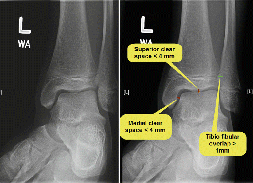
Fig. 22.4
Mortise view showing medial and superior clear space
Schuberth [42] showed that medial clear space was a poor predictor of arthroscopically-diagnosed deltoid ligament tears. They found the 88.5 % false positive rate for deltoid ligament rupture when medial clear space > or = 3 mm (P = .54, Fisher’s exact test) and 53.6 % when medial clear space > or = 4 mm (P = .007).
Several studies [41, 43, 44] have suggested that external rotation stress (manual or gravitational) x-ray can differentiate stable from unstable undisplaced fractures when plain x-rays are inconclusive. These studies assumed ankle instability when the clear medial space increased more than 4 mm with stress. However, this has been contested. Koval [41] investigated the significance of positive stress radiographs (medial clear space >5 mm on stress testing) in 21 patients who. All had MRI scan; 19 patients had evidence of partially torn deep deltoid ligament and their ankles were not stabilised whereas two had a complete rupture of the deep deltoid ligament and underwent stabilisation. All fractures united without evidence of residual medial clear space widening or posttraumatic joint space narrowing.
Several other studies concluded that stress radiographs greatly over-estimate instability and that most undisplaced ankle fractures can be treated non-surgically [45–47]. Most of these studies include only a few children and adolescents so their findings may not be applicable to paediatric practice.
The Role of CT Scan
CT can be useful in assessing more complex ankle fractures for accurate diagnosis, preoperative planning, and assessment of reduction of intra-articular fractures. However, clear indications are still lacking and reasoning remains subjective. A recent study of 64 distal tibial fractures with intra-articular involvement demonstrated the importance of CT scan in assessment [48]. All patients had plain radiographs and CT scans. Findings based on radiographs and CT scan are summarised in Table 22.4. Authors concluded that CT scan led to changes in fracture classification and treatment decision.
Table 22.4
The difference in classification and decisions of surgical treatment when CT scan was used in 64 patients with ankle fracture
Palin radiograph | Adding CT Scan | |
|---|---|---|
SH-III fracture | 31 | 20 |
SH-IV fracture | 8 | 12 |
Tillaux fracture | 9 | 9 |
Triplane fracture | 16 | 23 |
Decision for operative treatment | 18 | 42 |
Decision for non operative | 46 | 22 |
In a similar study of 25 triplane fractures by Eismann [49], there was poor inter-rater reliability (a kappa of 0.17) and intra-rater reliability (a kappa of 0.31) with radiographs alone but moderate inter-rater reliability (a kappa of 0.41) and intra-rater reliability (a kappa of 0.54) with the addition of computed tomography. More interestingly, the decision from non-operative to operative treatments was changed in 27 % and either the orientation or number of screws was changed in 41 % of the cases when raters reviewed CT scans.
Black et al. [50] performed a retrospective analysis on 100 consecutive patients treated for ankle fractures having both preoperative radiographs and CT scans. They found operative strategy was changed in 24 % of cases after CT review. Several authors suggested that CT scan should be routinely performed in clinical practice [51, 52].
The Role of Magnetic Resonance Imaging (MRI)
MRI provides fine detail of bones and soft-tissue structures and does not involve radiation. MRI scans have been used to diagnose occult fractures and, in cases of incomplete ossification of malleoli, to establish displacement [41, 53].
Lohman et al. [53] studied 60 children with acute ankle injuries with both conventional radiography and MR. Plain radiography produced 5 of 28 (18 %) false negative and 12 of 92 (13 %) false positive fracture diagnoses compared to MRI. However they found no complex ankle fracture was missed and the MRI did not change the treatment plan in any case.
Petit et al. [54] compared radiographs with MRIs for 29 children with physeal ankle injuries and found only 1 of 29 fractures (3 %) was misclassified by plain film radiography.
Contrary to these two studies, Carey et al. [55] reported on 14 patients with suspected physeal injuries and indicated that that the MRI scans led to a change in the Salter-Harris classification for two of the nine patients with fractures seen on radiographs, they identified two occult fractures, and changed the management in five patients.
What Is the Best Treatment for Non-displaced Ankle Fractures in Children?
There is a consensus on non operative treatment of stable and undisplaced ankle fractures. Many centres use a below knee full cast to treat undisplaced stable fractures although this may not be necessary in every case. In a randomised controlled study (level I) of 40 patients with Lauge-Hansen supination-eversion, stage II ankle fractures, the use of the air stirrup led to a significant improvement in early patient comfort, post-fracture swelling, range of ankle motion at union, and time to full rehabilitation in comparison to a below knee walking cast [56].Similar findings were found in 66 patients who were treated with Aircast Stirrup ankle braces or Don Joy R.O.M.-Walker braces [57]. Subjective satisfaction with comfort and ease of use was significantly higher with Aircast Stirrup while pain relief and an inflammatory score were significantly better in the R.O.M.-Walker group after 4 weeks. There was in difference in outcomes after 3 months.
Related posts:
 Evidence-Based Treatment of Flexible Flat Foot in Children
Evidence-Based Treatment of Glenohumeral Dysplasia Caused by Obstetric Brachial Plexus Injuries
Evidence-Based Treatment of Flexible Flat Foot in Children
Evidence-Based Treatment of Glenohumeral Dysplasia Caused by Obstetric Brachial Plexus Injuries
 Evidence-Based Treatment of Deformity in Multiple Osteochondromatosis
 Evidence-Based Treatment for Slipped Upper Femoral Epiphysis
 Private: Physeal Injury, Epiphysiodesis and Guided Growth
 Femoro-Acetabular Impingement in Children
Stay updated, free articles. Join our Telegram channel

Full access? Get Clinical Tree


