I Foundations and Techniques 1 Visceral Manipulation according to Barral 2 Fascial Treatment of the Organs according to Finet and Williame 3 Circulatory Techniques according to Kuchera 4 Reflex Point Treatment according to Chapman We distinguish three movements of the internal organs: motricity, mobility, and motility. Motricity refers to passive changes in the position of the organs that result from arbitrary motor activity by the locomotor system. If, for example, you bend the upper body to the right, the move compresses the abdominal organs on the right side but stretches the wall of the torso on the left, resulting in a pull on the left-sided organ attachments which enlarges their available space. When bending the upper body forward, the intraperitoneal organs migrate anteriorly as the result of gravity and their high degree of mobility. Any activity that involves continuous sitting compresses the small and large intestines and impairs their peristalsis. Lifting both arms in maximum flexion results in an extension of the thoracic spinal column (TSC) and an inspiration position of the ribs. As the parietal pleura follows this movement of the thorax, and the lung is connected to the movement of the chest by its stretch, the lung increases its volume without having to make any additional respiratory effort. In visceral manipulation, mobility refers to the movement either between two organs or between an organ and the wall of the torso, the diaphragm, or another structure in the musculoskeletal system. The engine for this movement can be motricity or different “automatisms.” Automatism refers to a movement that is performed involuntarily by striated or smooth muscles. Furthermore we can differentiate between automatisms that occur continuously and movements of the organs marked by periodicity. Automatisms include: Diaphragmatic breathing. With 12–14 breaths/min, the diaphragm contracts about 20000 times a day. In doing so, it acts like a piston sliding up and down in a cylinder. During inspiration, the diaphragm sinks caudally, the volume of the thorax increases, and the abdominal organs migrate downward. The soft muscular abdominal wall allows the abdominal organs to move anteriorly out of the way; as a result, the volume of the abdomen hardly changes at all during inspiration. During expiration, the opposite movement occurs. Heart action. At 70 heartbeats/min, the heart contracts about 100000 times a day. These actions act like vibrations on the mediastinal organs and, via the diaphragm, also on the abdomen. Motility is defined as the intrinsic movement of the organs with a slow frequency and small amplitude. It can be detected by the hand of a trained practitioner and is the kinetic expression of movements in the organ tissues. During embryonic development, the evolving organs carry out growth movements and position shifts that remain stored in each organ cell as a kind of memory. Motility is a rhythmic repetition of this embryonic migration to its place of origin and back to the final, postnatal position. Likewise, it is impossible to rule out a connection to the craniosacral rhythm, in spite of the fact that motility shows a different frequency. We distinguish between a so-called expiration phase, that is, the movement toward the median line, and an inspiration phase, a movement in the opposite direction away from the median line. The frequency is 7–8 cycles/min, one cycle comprising one expiration and one inspiration. Motricity, automatisms, and mobility cause changes in positional relationships of the organs. The movement occurs along a defined axis with a defined amplitude and, thereby, the organs with structural relationships to each other act in a similar way to a joint in the locomotor system. Two joint partners form the visceral joint; the two joint partners can be two organs (liver–kidney) or an organ and a muscular wall (liver–diaphragm). The serous membranes—pleura, peritoneum, pericardium, and meninges/peripheral nerve sheaths—constitute most of these gliding surfaces. The joint partners are fixed to each other: there are several attachments on the organs that are important for the axis of movement—see box. Wherever we find a film of fluid (peritoneum, pleura, pericardium), the organs of a visceral joint are both separated from each other and connected by this fluid. They act in a similar way to two panes of glass with a drop of fluid between them—they can glide past each other, but the adhesive force keeps them together. In visceral manipulation, ligaments are pleural or peritoneal folds that connect an organ to either the wall of the trunk or other organs. In most cases, they do not contain blood vessels but are sensitive and well innervated. They fix the organs against gravity. Turgor or intravisceral pressure refers to the ability of an organ to occupy the largest space possible. The reasons for this characteristic are elasticity, vascular effects (decreased or increased blood circulation), and gases in hollow organs. The intracavitary pressure is the sum of all intravis-ceral pressures plus the pressure between the organs. This pressure causes the organs to be pressed and fixed against each other. As a result, we find a large excess of pressure in the abdomen, which is countered by a vacuum in the thorax. The diaphragm is the border layer between these pressure states. The organs near the diaphragm are influenced greatly by pressures. A diaphragmatic hernia will thus always lead to a movement of organ parts from the abdomen into the thorax, against gravity. This illustrates the great potency of such pressure effects on the fixation of the organs. The mesenteries are duplicatures the peritoneum with only a minor role in fixation. They supply the organ’s blood circulation. The omenta are also infoldings of the peritoneum that connect two organs to each other. Their role in organ fixation is rather small, although their vasculonervous function is of more importance. Organs move around specific axes and with defined amplitudes. Changes in the axes of movement or amplitudes lead to deviations from the physiologic mobility or motility. Such changes lead to In principle, we distinguish between disturbed mobility and disturbed motility. An organ completely or partly loses its ability to move as a result of the following causes. Articular restrictions. This dysfunction can lead to disturbed mobility and disturbed motility. If only the motility but not the mobility is disturbed, we speak of “adhesions.” If, however, both movement qualities are impaired, we call this “fixations.” In fixations, the axis of movement and the amplitude could have changed. Causes include: As a result, we notice a change in motility, especially in amplitude. Altered mobility affects the organ only when the viscerospasm has also adversely impacted the organ attachments. Causes for irritations include: Loss of ligamentary elasticity (ptosis). The loss of elasticity in ligamentary attachments causes diverse organs, such as the transverse colon, kidney, or urinary bladder, to descend with gravity. The axes and amplitude of mobility change, as does motility, the causes of which include: Motility can have its amplitude disturbed. The range of motion can be reduced in either one direction or both directions. A disturbance also alters the rhythm of the movement: The causes include: Questioning the patient allows the practitioner to collect information about the following keywords: During an osteopathic inspection with the patient in the standing position, the following should be noted: Even this long list is not exhaustive. Ultimately, we look for findings that guide the practitioner to the dysfunctional organ or into the diagnostic zone. In visceral manipulation, for example, we interpret posture abnormalities in light of the fact that the body creates convexities to compensate by allowing organs more room and concavities to compensate by providing protection to underlying structures. An upper abdomen that protrudes into the epigastric angle indicates dysfunction in the upper abdominal organs, which need space and move away anteriorly. When palpating this region, we are almost certain to find pain in individual organs, such as the stomach, or to trigger symptoms such as nausea. A left convex scoliosis with the vertex point of the concave curvature in the area of the right lower costal arch can indicate dysfunction in the liver or gallbladder. Compression of the organ by the concavity reduces mobility and provides rest or immobilization. This mechanism is comparable to a parietal joint that stops hurting when the person no longer moves it. In palpation of the thorax, elasticity tests are performed at different locations on the ribs and sternum, to gain an impression of the fascial tensions in the ribcage. Abdominal palpation is accomplished in two steps. During superficial palpation of the abdomen, the various regions of the abdomen (epigastrium, hypochondrium, etc.) are palpated with both hands in the fascial plane. This noteworthy layer consists of the fascia of the abdominal muscles, the greater omentum, and the parietal anterior peritoneum. To reach it in palpation, sink both hands into the abdomen until you detect the organs under your fingers. Then take the pressure off the abdomen just to the point where you no longer detect the organs. Evaluate the patient for differences in tension between the two sides, triggering of pain by the palpation, and possibly evaluate existing scars for tension and sensitivity. Deep palpation is applied to the organs themselves. We evaluate for: Be particularly mindful of whether the palpation could cause parietal symptoms such as lumbalgia. This would indicate a possible causal link to the palpated organ. In addition, pay attention to vegetative symptoms that can be triggered by the examination: These symptoms can be signs of acute disorders (e.g., cholecystitis) which are a contraindication to osteopathic treatment.
1 Visceral Manipulation according to Barral
Theory of Visceral Manipulation
 Physiology of Organ Movement
Motricity
Mobility
 diaphragmatic breathing
 heart action
 peristalsis of the visceral hollow organs in the gastrointestinal tract
Motility
 Visceral Joint
Double-Leaf System
Ligamentary System
Turgor and Intracavitary Pressure
Mesenteries
Omenta
 Pathology of Organ Movement
 local pathologies first without and later with symptoms
 recurring local pathologies
 pathologies in visceral or parietal regions of the body that are linked via topographic, vascular, nervous, or fascial osteopathic chains
Disturbed Mobility
 infections inflammation
 surgical interventions
 blunt trauma
 inflammation
 vegetative dysinnervation
 allergic reactions
 psychosomatic influences
 a result of adhesions
 asthenic constitution
 anorexia or rapid weight loss due to other causes
 age-related loss of elasticity
 depression with generalized tonus reduction
 general laxity at the end of or after pregnancy
 delivery by vacuum extraction
 multiparity
Disturbed Motility
 The rest phase between inspiration and expiration can be prolonged.
 We detect an arrhythmic motion.
 The frequency is reduced.
 general loss of vitality in the organ as a sign of pathology
 articular restriction
 ptosis
 viscerospasm
Diagnosis and General Treatment Principles in Visceral Osteopathy
 Medical History
 current reason for consultation
 patient history with chronological list of items, for example:
 gynecologic history in women, for example:
 urologic history in men:
 Inspection
Example of a Convexity
Example of a Concavity
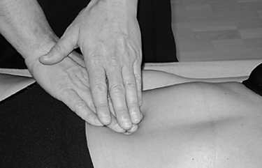
 Palpation
Superficial Palpation
Deep palpation
 painfulness
 differences in tension
 position of the organ
 tone of the organ
 nausea and vomiting
 sweating
 tachycardia
 tendency to collapse
 dizziness
 severe pain leading to tension that actively resists the palpation
Related posts:

Stay updated, free articles. Join our Telegram channel

Full access? Get Clinical Tree


