Current Concepts and Techniques in Foot and Ankle Surgery
Pigmented Villonodular Synovitis of the Distal Tibiofibular Joint: A Case Report
Keywords
• Pigmented villonodular synovitis • Distal tibiofibular joint • Surgical treatment
Pigmented villonodular synovitis (PVNS) is a rare proliferative disorder of the synovium that affects young and middle-aged adults.1–6 PVNS is usually monoarticular and often arises in the joints.5 This condition may be locally destructive and involve muscles, tendons, bursae, bone, and skin.6 The most common locations are the knee and hip, flexor tendon sheaths of the hand, ankle, and shoulder joints. The sternoclavicular and tibiofibular joints are rare locations.7–10 Patients frequently present with pain, joint effusions, and swelling. The duration of symptoms varies.5,11
Plain radiographs may show defects in the adjacent bone, bone cysts, and a sclerotic rim.12,13 Effusions may appear dense on plain radiographs and computed tomographic (CT) images because of high iron content. Magnetic resonance imaging (MRI) is useful to delineate the soft tissue extent and bone involvement, if present, of PVNS, showing low signal intensity on both T1- and T2-weighted images.3,6 The extent of the abnormality may seem larger than it actually is because of the blooming phenomenon of the magnetic susceptibility effect caused by hemosiderin.6,10 Macroscopically, PVNS appears as thickened reddish-brown synovium (because of hemosiderin deposition) with numerous villous projections. Microscopically, the lesions are composed of matted villi containing thin-walled vascular channels and supporting stroma closely packed with polyhedral stromal cells, multinucleate giant cells, and macrophages that may contain hemosiderin and lipids. Abundant collagen, fibrosis, and hyalinization may be present in patients with long-standing PVNS.1,2,6
The incidence of PVNS in the ankle joint is approximately 2.5%,11 but the occurrence in the distal tibiofibular joint is relatively rare.14 This case report presents a patient with PVNS of the distal tibiofibular joint. To the best of the authors’ knowledge, this case is only the second report in the English language medical literature of isolated involvement of the distal tibiofibular joint.
Case report
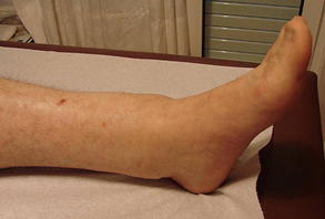
A 40-year-old man presented with a 3 months’ history of edema of the left distal tibia and ankle (Fig. 1). His past medical history was unremarkable. The patient denied any previous trauma to the area. The results of the laboratory examinations were within normal values.
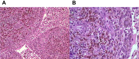
Radiographs of the left distal tibia and ankle joint showed irregularity of the distal tibiofibular joint (Fig. 2). The CT scan results showed erosion of the distal tibiofibular joint and a large soft tissue mass extending through both sides of the joint (Fig. 3). The MRI results showed a soft tissue mass with a maximal diameter of 95 mm, extending through the distal tibiofibular joint to the anterior and posterior compartments of the distal tibia with erosion of the tibia and fibula (Fig. 4). The bone scan results showed increased radioisotope uptake at the left distal tibia (Fig. 5). Histologic sections of the biopsy specimens showed proliferation of round to polygonal cells, with scant cytoplasm forming villous and fingerlike or rounded mass underlying the synovium. Multinucleated giant cells were scattered throughout the lesion. Hemosiderin-laden macrophages varied in number, giving the synovium a dark-brown appearance (Fig. 6). These findings were consistent with the diagnosis of PVNS.
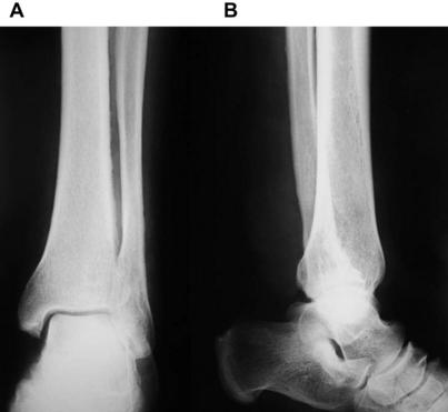
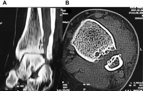
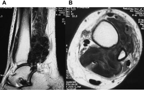
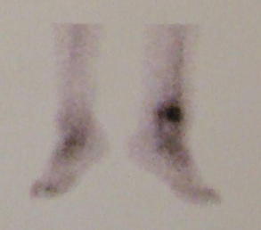
Fig. 5 Technetium Tc 99m bone scan image shows increased radioisotope uptake in the left distal tibia.
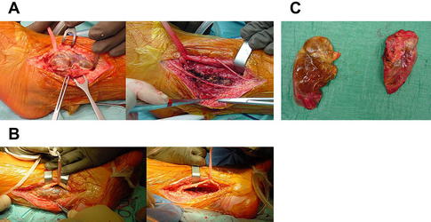
Through the anterolateral (Fig. 7A) and posteromedial (see Fig. 7B) approaches, complete excision of the proliferative synovium was done (see Fig. 7C). Histologic evaluation of the excised specimen was consistent with the biopsy result. Postoperative recovery of the patient was uneventful. At the latest examination, 3 years after diagnosis and surgical treatment, MRI results showed no evidence of local recurrence (Fig. 8).


Stay updated, free articles. Join our Telegram channel

Full access? Get Clinical Tree


