9 Vertebral column
The vertebral column usually consists of:
7 cervical vertebrae (movable)
12 thoracic vertebrae (movable)
4 coccygeal segments (fused to a variable extent).
A ‘typical’ vertebra
Main parts
Inferior articular processes –
projections on the inferior aspect of the vertebral arch which carry the inferior articular facets.
Vertebral notches, superior and inferior –
formed between the body and the articular processes above and below the pedicles.
Cervical vertebrae
3rd to 6th (typical) (Figs 9.1 and 9.2)
Features
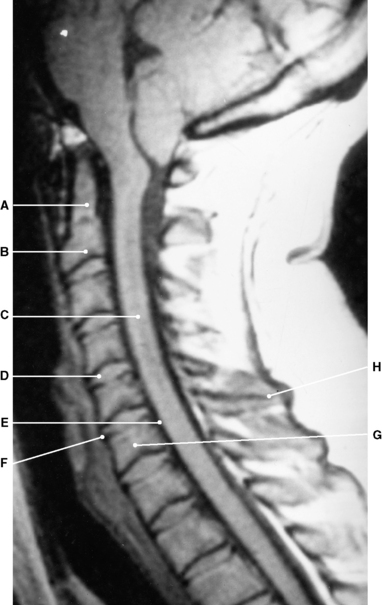
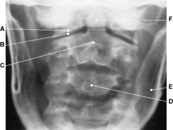
Radiographic appearances of the cervical vertebrae (Figs 9.3, 9.4, 9.5, 9.6 and Plate 6)
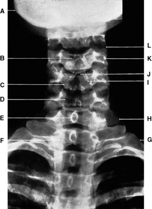
Fig. 9.3 Third to seventh cervical vertebrae: anteroposterior projection.
C–Body of 5th cervical vertebra
E–Spinous process of 7th cervical vertebra
F–Body of 1st thoracic vertebra
H–Transverse process of 7th cervical vertebra
(From Bryan 1996.)
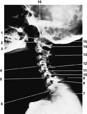
Fig. 9.4 Cervical vertebrae: lateral projection.
1 – Anterior tubercle of atlas
4 – Body of 4th cervical vertebra
6 – Body of 1st thoracic vertebra
9 – Spinous process of 7th cervical vertebra (vertebra prominens)
10 – Inferior articular process
11 – Superior articular process
14 – Posterior tubercle of atlas
(From Bryan 1996.)
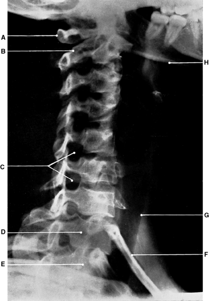
Fig. 9.5 Cervical vertebrae: oblique anteroposterior projection.
C – Right intervertebral foramina
(From Bryan 1996.)
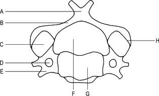
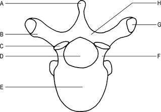
1st cervical vertebra (atlas) (Fig. 9.7)
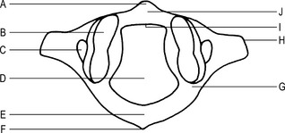
Fig. 9.7 1st cervical vertebra (superior aspect).
G–Groove for the vertebral artery
I–Facet for the odontoid process
Articulations
Superior articular facets with the occipital condyles to form the atlanto-occipital joints.
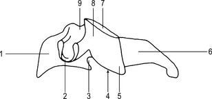
2nd cervical vertebra (axis) (Figs 9.8 and 9.9)
Articulations
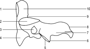
Fig. 9.9 2nd cervical vertebra (axis) (lateral aspect).
1 – Facet for the anterior arch of the atlas
6 – Inferior articular process
9 – Groove for the transverse ligament of the atlas
The body with the body of the 3rd cervical vertebra to form the intervertebral joint.
Ossification
Primary centres
odontoid process – 2 centres appear 6th month intrauterine life.
odontoid centres fuse together before birth;
vertebral arch – 2 centres appear 7th–8th month intrauterine life;
Thoracic vertebrae
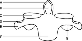
2nd to 8th (typical) (Figs 9.13 and 9.14)
Features
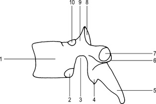
Fig. 9.14 A typical thoracic vertebra (lateral aspect).
2 – Demi-facet for the head of the rib
4 – Inferior articular process
7 – Costal facet for the tubercle of the rib
9 – Superior articular process

