Vascularized Bone Grafting for Kienböck Disease

56 Vascularized Bone Grafting for Kienböck Disease


Indications


Vascularized bone grafts (VBGs) can be used for revascularization in early or advanced Kienböck disease with an ulnar neutral or plus variance as long as the cartilage shell is intact and no arthrosis is found. VBGs can also be used as an adjunctive procedure to radial shortening in patients with an ulnar minus variance. This technique is not indicated when there is fragmentation of the lunate or arthritis.


Technique


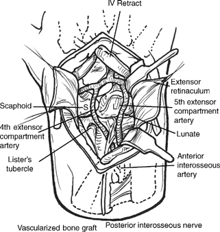
  • Longitudinal dorsal incision that begins just ulnar to Lister’s tubercle and extends to the base of the third metacarpal (Fig. 56-1)
  • Protect the radial sensory nerves and lateral antebrachial cutaneous branches using loupe magnification. Elevate flaps at the level of the extensor retinaculum.
  • The extensor digitorum communis compartment is identified and opened. The fourth extensor compartment artery (ECA) is identified deep to the tendons along the radial aspect. The fourth ECA is adjacent to the posterior interosseous nerve (PIN) (Fig. 56-1).
  • The fourth ECA is dissected in a proximal direction to its origin from the posterior division of the anterior interosseous artery (AIA). At this site, the fifth ECA is located as it connects to the same division. The fifth ECA is dissected in a distal direction toward the dorsal intercarpal arch.
  • The anterior interosseous artery is ligated proximal to the origins of the fourth and fifth ECAs to create a single pedicle (Fig. 56-2).
  • A VBG is configured 1 cm proximal to the radiocarpal joint and centered over the fourth ECA. The
  • VBG is carefully harvested using small curved and straight osteotomes. The vascular pedicle is protected. The VBG is elevated on its common pedicle and retracted in an ulnar direction (Fig. 56-2).
  • A longitudinal arthrotomy is performed over the lunate, which is inspected for shape and configuration. The cartilage shell of the lunate should be intact to accept a VBG. The shell must be preserved during manipulation of the lunate. The necrotic bone is removed using curettes via a dorsal window (Fig. 56-3). Any collapse is gently expanded in a sequential fashion.
  • Additional cancellous bone graft is harvested from the distal radius using the cortical defect created by the harvest of the VBG. The bone graft is packed within the lunate.
  • The tourniquet is deflated to verify blood flow with the VBG. The VBG is fashioned to wedge within the lunate and dorsal window. The cortical surface serves as a strut across the reconstituted lunate. The pedicle is protected during the insertion of the VBG (Fig. 56-4). Internal fixation is not used.
  • An external fixator is applied to bridge the radiocarpal joint and unload the lunate during revascularization. Pins are inserted into the second metacarpal and radius using small incisions. A small amount of distraction is applied.

image

Only gold members can continue reading. Log In or Register to continue

Stay updated, free articles. Join our Telegram channel

Mar 21, 2017 | Posted by in MUSCULOSKELETAL MEDICINE | Comments Off on Vascularized Bone Grafting for Kienböck Disease

Full access? Get Clinical Tree

Get Clinical Tree app for offline access