Fig. 10.1
Anteroposterior and lateral X-rays of valgus knee demonstrating the biplanar (frontal and sagittal) bone wear of the lateral tibial plateau (red dotted lines) with distraction of the medial side (yellow arrow)
External rotation of the tibia in relation to the femur has an important influence on the femoropatellar joint, increasing the Q-angle and lateral displacement forces on the patella, which overloads its lateral side and negatively impacts patellar tracking, leading even to dislocation.
The structural changes described above must be taken into account during bone resection, ligament balancing, and rotational positioning of implants during TKA, in order to avoid unsatisfactory limb alignment, patellar maltracking, or knee instability (Fig. 10.2).

Fig. 10.2
Case of a female patient with severe valgus knee, reconstruction with augment of the lateral tibial plateau, anterior tibial tuberosity osteotomy, and release of the lateral structures to achieve soft tissue balance without the need for a constrained implant
Valgus deformity may be constitutional or acquired.
Constitutional deformity is most frequently bilateral, with an extra-articular origin of the deformity, usually on the femoral side. In cases of significant tibial or femoral contribution to valgus deformity, correctional osteotomy should be considered.
Acquired valgus may be secondary to primary osteoarthritis, rheumatoid arthritis, metabolic disorders such as rickets or renal dystrophy, osteonecrosis, and overcorrection after proximal tibial osteotomy. There are also post-traumatic cases resulting mostly from malunion of tibial plateau fractures.
10.3 Anatomy of the Lateral Compartment of the Knee
The ligaments, capsules, and muscles all contribute to stability of the lateral compartment. The ligamento-capsular elements are represented by the lateral collateral ligament (LCL) and the posterolateral angle complex. The muscles of the lateral aspect of the knee can be divided into anterolateral (iliotibial band (ITB)) and posterolateral groups (the popliteus, biceps femoris, lateral head of the gastrocnemius) (Fig. 10.3).

Fig. 10.3
Schematic representation of lateral knee anatomy
The ITB inserts principally onto Gerdy’s tubercle, but it runs also toward the patella as the iliopatellar band and to the lateral intermuscular septum, fibula, and biceps femoris. The ITB is the lateral stabilizer of the first 30° of knee flexion, acting against varus deformation forces, and also has a role in stabilizing internal rotation during knee flexion [4].
The LCL runs from the lateral epicondyle of the distal femur to the anterolateral surface of fibular head and acts between 0° and 90° of knee flexion.
The posterolateral angle complex is a musculoligamentous unit consisting of the popliteus tendon (POP) and the elements reinforcing the posterolateral capsule (PLC): the oblique popliteal ligament (OPL), arcuate ligament, popliteofibular ligament, and fabellofibular ligament. All stabilize the knee between 0° and 30° of flexion.
The popliteus muscle tendon runs obliquely, passing under the LCL to insert anterior to it on the lateral femoral condyle. It is active between 60° and 90° of flexion and its contraction causes internal tibial rotation [5].
The biceps femoris is an important landmark for the common peroneal nerve, which is at risk during extensive lateral release [6, 7] or release of biceps femoris from the proximal fibula.
The posterior articular capsule works in extension and is located just in front of the lateral gastrocnemius. It may be released in a subperiosteal manner in cases of fixed flexion.
To simplify, the lateral elements can be divided into two groups: those inserting near the transepicondylar axis (LCL and POP), important in both extension and flexion, and those inserting at more distally (ITB, posterolateral capsule, biceps, and lateral gastrocnemius), acting only in extension [8].
Isolated sectioning of one of the aforementioned structures does not destabilize the lateral compartment, and differential opening of the lateral compartment is observed, most notably after LCL section. However, extensive release by sectioning of all these elements will cause the substantial widening of lateral space, further increased if the posterior cruciate ligament (PCL) is sacrificed [8–11]. This effect is more significant in flexion than in extension, making balancing of the resulting asymmetric gaps complicated.
Lateral femoral condyle hypoplasia is the subject of controversy. Brihault et al. [12] demonstrated the predominant importance of femoral valgus over hypoplasia. However, the reduced size of the condyle has implications for “filling in” the lateral space with a more bulky implant and overstuffing. As a consequence it implies closure of the capsular plane problematic during lateral approach (Fig. 10.4).

Fig. 10.4
Lateral condyle modification in valgus knee with hypoplasia or wear and the implication for femoral implant placement
10.4 Radiological Assessment of the Arthritic Valgus Knee
Radiological diagnosis of the valgus knee is based on weight-bearing anteroposterior radiographs in extension and lateral radiographs in 30° of flexion, Rosenberg (Schuss) views for femorotibial space assessment, and weight-bearing full-length radiographs for HKA angle evaluation. This last view also permits determination of the femoral and tibial mechanical axes and precise determination of the origin of the valgus deformity. Significant flexion deformity or lower extremity rotational deformities may cause error in HKA angle measurement. It has been shown, however, that rotation of up to 20° has little effect on HKA [13]. Stress valgus-varus radiographs should be performed to evaluate reducibility of the deformation and to judge any associated degree of ligament laxity. Lateral radiographs show the tibial slope. Coexisting patellofemoral OA is evaluated on the patella skyline (Merchant) view at 30° of flexion (Fig. 10.5). The frequent lateral subluxation of the patella, and its diminished thickness in the advanced stages, can be appreciated, influencing the decision for patella resurfacing.

Fig. 10.5
Full set of radiographs required for evaluation and planning TKA in the valgus knee
The radiological evaluation helps moreover to appreciate the distribution and amount of osteophytes and loose bodies, the presence of extra-articular deformities or pathologies, and the general bone quality.
10.5 General Considerations in TKA Management in Valgus Knee
When planning TKA in valgus knee, the surgeon must make several decisions concerning:
The approach, being medial or lateral, and the requirement for tibial tubercle osteotomy
The amount and order of soft tissue releases and the requirement for condylar osteotomy
Level of implant constraint
Extra-articular deformity correction, either simultaneous or two stage
Many factors influence these decisions, including the magnitude and location of deformity, the degree of rigidity, the presence of medial laxity, the amount of bone loss, the preexisting knee flexion deformity, and the condition of the patellofemoral joint.
According to Krackow et al. [10], at least three situations can be apparent (Fig. 10.6):

Fig. 10.6
Classification of arthritic valgus knee: Stage I – wear of lateral compartment with or without the tightness of lateral structures. Stage II – wear of the lateral compartment with laxity of the medial side. Stage III – extra-articular deformity (e.g., after valgus-producing osteotomy)
First – isolated wear of the lateral femorotibial compartment, reducible or not, with competent medial structures
Second – lateral bone wear associated with medial laxity
Third – lateral bone wear combined with femoral or tibial extra-articular deformity (previous osteotomy or post-traumatic malunion)
10.6 Approach Choice
The choice of approach remains controversial. The surgery can be done by either a lateral or medial approach.
10.6.1 Medial Approach
The medial approach is advantageous in that it is the most commonly used and well-known approach in arthroplasty surgery of the knee. It is easy to perform in valgus knee without tight lateral structures and where there is no flexion deformity. Its usefulness in the valgus knee is improved by the technique of intra-articular or “inside-out” lateral structure release.
It is well described by Ranawat et al. [1]. The medial parapatellar arthrotomy is performed followed by minimal subperiosteal liberation of the medial structures just to allow knee exposure and resection of cruciate ligaments and the menisci.
The osteophytes are removed and the tibial cut is made perpendicular to its anatomical axis. The thickness of resected bone of the unaffected medial part should not exceed 6–8 mm.
The femoral distal cut is performed with the use of an intramedullary guide. The femoral valgus angle is decreased from usual 6–3°, which results in a varus cut of the distal femur. This cut is also minimized and should measure no more than 10 mm on the medial condyle, resulting in a thin or almost no lateral condyle cut.
Ligament balancing is performed firstly in extension by decreasing the tightness of the lateral structures as required in a stepwise manner. The order and degree of release vary according to different authors [1, 9–11, 14–16]. The sequence proposed by Ranawat et al. [1] is the one mostly employed.
The tibiofemoral space is opened in extension with the Meary’s spreader. This allows palpation and identification of the tight structures: the posterolateral angle, ITB, and LCL.
Release begins in general with the posterolateral capsule, which is cut at the level of the tibial osteotomy between the posterior border of the ITB and the POP, followed by pie crusting of the capsule above the initial cut (Fig. 10.7). It is at that moment the LCL can be elongated according to Elkus et al. [16]. The POP is preserved unless it is too tight. The ITB is elongated with the pie-crusting technique or is liberated from Gerdy’s tubercle as required.
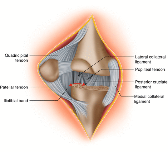
Fig. 10.7
Schematic representation of the right knee with selected structures seen via a medial parapatellar approach, with indication of section level (red arrow) of the lateral elements after femoral and tibial cuts. MCL medial collateral ligament, PCL posterior cruciate ligament, POP popliteal tendon, LCL lateral collateral ligament, ITB iliotibial band, QT quadricipital tendon, PT patellar tendon
At least one lateral structure should be preserved in order to maintain lateral stability. If after ligament balancing in extension there is more than 5 mm of mediolateral laxity, a more constrained prosthesis should be used.
The flexion gap is balanced by adapting the thickness of the posterior femoral condyle cut and rotation of the femoral implant. The knee is flexed to 90° and distracted, and the femoral cutting guide is placed in such a way that its posterior edge is parallel to the tibial cut. The observed asymmetry of the posterior condyle cut creates a systemic gap. To achieve appropriate femoral rotation, in addition to using Whiteside’s line as reference, the transepicondylar axis and adequate tensioning in flexion of both compartments should be used [16]. The use of the posterior bicondylar line as reference runs the risk of placement of the femoral implant in internal rotation.
Down- or upsizing of the femoral implant can help in obtaining equality of both spaces. When mediolateral stability in different degrees of flexion is achieved, and after verifying the absence of notching of the anterior femoral cortex, the cutting guide can be fixed and final cuts performed.
The advantage of the medial approach is the choice of which structures are released and in which sequence. The disadvantages are related to weakening of the medial structures and limited access to posterolateral side due to the lateral subluxation of the extensor apparatus, which increases external rotation of the tibia. However, the sliding lateral condyle osteotomy (SLCO) is possible through this approach [17]. Access to the medial structures is easy if medial condyle osteotomy or tensioning of MCL is required.
10.6.2 Lateral Approach
The lateral approach is confusing and more difficult because of anatomical landmark inversion and different management of the soft tissues and the patella. It is, however, logical, giving direct access to the tight lateral structures and conserving the medial structures. It does not allow for tensioning procedures of the MCL [18], which are nonetheless exceptional. On the other hand, it facilitates lateral procedures such as the SLCO [12].
This approach, popularized by Keblish et al. [19] and Buechel et al. [7], follows the lateral border of patellar tendon in its distal part. Keblish proposed a lateral parapatellar arthrotomy with plasty of the lateral femoropatellar retinaculum and the use of the retropatellar fat pad during closure. Mertl et al. [20] pointed out the utility of tubercle osteotomy.
The skin incision is midline, centered on the patella, or slightly eccentric, following its lateral border. The transquadricipital arthrotomy starts proximally between the vastus lateralis and rectus femoris muscles and runs distally to the superolateral corner of the patella. Sectioning of the lateral retinaculum is done in a “Z-plasty” fashion, by separating the retinacular and capsular layers. The retinaculum is sectioned about 3 cm laterally from the patella’s edge, whereas the capsule is cut just next to it [19]. The retropatellar fat pad is detached from the patellar tendon and attached to the lateral capsular flap. This allows expansion of the lateral capsular layer and facilitates closure [7, 19]. The capsule is incised distally up to Gerdy’s tubercle. The ITB is detached, maintaining continuity with the anterior sural aponeurosis, and soft tissue subperiosteal detachment is continued to the proximal tibiofibular articulation. Special attention must be paid to the common peroneal nerve. In this manner the ITB is loosened, and lateral structure (LCL, POP, and posterolateral capsule) release may be performed depending on the degree of tightness of the involved structures (Fig. 10.8). Initial simultaneous release of both the LCL and POP should be avoided as it results in significant laxity. It may, however, be indispensable in cases with significant flexion deformity.

Fig. 10.8
Stage I – wear of the lateral compartment with tightness of lateral structures, release through the lateral approach starting with ITB release, following by the more posterior elements (LCL and/or POP), eventual section of PCL
In general, the patella is inverted, rarely dislocated. In the situation of patella baja or the danger of distal peel off of extensor apparatus, a tibial tubercle osteotomy or detensioning of the quadricipital tendon by the “rectus snip” technique should be considered. A tibial tubercle osteotomy is preferred to patella tendon detachment, which has an uncertain prognosis.
Related posts:
 Soft Tissue Balance of the Native Knee Provides Guidance for Balancing a Total Knee Arthroplasty
Anatomy and Biomechanics of the Native Knee and Its Relevance for Total Knee Replacement
Assessment in Primary TKA: Intraoperative Assessment Tensor
Trouble Shooting: Intraoperative MCL Injury
PS: Gap Technique
Trouble Shooting: Intraoperative MCL Injury
Soft Tissue Balance of the Native Knee Provides Guidance for Balancing a Total Knee Arthroplasty
Anatomy and Biomechanics of the Native Knee and Its Relevance for Total Knee Replacement
Assessment in Primary TKA: Intraoperative Assessment Tensor
Trouble Shooting: Intraoperative MCL Injury
PS: Gap Technique
Trouble Shooting: Intraoperative MCL Injury
Stay updated, free articles. Join our Telegram channel

Full access? Get Clinical Tree


