Undisplaced Scaphoid Fractures
9.1 Introduction
A broken scaphoid represents 2% to 7%1 of all fractures, and between 88% and 90% will unite if immobilized in a cast. However, 10% to 12% of scaphoid fractures do not unite, with a higher incidence (14—50%) in displaced fractures.2 If a scaphoid fracture nonunion is left untreated, the wrist becomes arthritic, usually within 5 years, and after 10 years all will have arthritis on radiographs. Scaphoid fracture affects patients at a very young age. That is why treating this fracture at the first instance is so important.
Systematic reviews3,4,5 have found little evidence from randomized controlled trials to inform treatment decisions for scaphoid waist fractures. It was not clear whether patients who had surgical fixation of undisplaced or minimally displaced scaphoid fractures had better long-term benefit than those treated in a cast. Surgery avoids a cast and helps early return to previous activity level and function, but can expose patients to a higher complication rate of between 9% and 22%, although complications are usually minor.6
The term “scaphoid” derives from the Greek word “skaphe” meaning a boat. This bone lies obliquely and links the distal row of the carpus to the proximal row. Forty-two percent of the scaphoid surface is articular and is covered with cartilage and 58% is nonarticulating. This means that most fractures of the scaphoid are bathed in synovial joint fluid and are intra-articular.
The radial artery supplies the scaphoid but the supply is precarious. Perforating branches along the dorsal scaphoid ridge supply 75% of the bone. These dorsal branches also supply the proximal pole in a retrograde manner. Obletz and Halbstein7 established that 67% of 297 cadaver scaphoids had multiple arterial foramina throughout their length; 13% had foramina only in the distal one-third, and 20% had arterial foramina near the waist of the scaphoid bone with often only a single foramen over the proximal part of the scaphoid. In such scaphoids a fracture could significantly disrupt circulation to the proximal pole and take much longer to heal.
Scaphoid fractures are common, especially in young men.8 These usually occur after a fall on the outstretched hand, and often during sporting activity.
The scaphoid breaks when there is forced extension of the wrist. This can happen when a ball strikes the palm of the hand, forcing the wrist back. It can also happen when a starting handle suddenly whips the hand back. Occasionally the scaphoid can break on forced palmar flexion of the wrist such as when being struck on the back of the hand. Studies trying to create scaphoid fractures in cadavers have failed to establish a consistent injury mechanism.
An undisplaced scaphoid fracture is defined as the one in which the fracture gap or step is less than 1 mm on a set of radiographic views. This chapter discusses such fractures and their management.
9.2 Epidemiology
Fracture of the scaphoid is the commonest fracture affecting bones of the carpus. Hove1 documented that 60% of fractures affecting carpal bones involved the scaphoid. The incidence established in Norway is far higher than that found in the United Kingdom of 29/100,000/year,9 or that in the United States, even in the young, fit, and active military population.10
9.3 Assessment
Patients complain of pain on the radial side of the wrist after an injury, such as a fall on the palm of the hand. The wrist is often not swollen nor is it significantly tender. These (typically) young men usually ignore the pain on the radial aspect of the wrist, treating it as a minor injury. It is not unusual for these patients to present a few days or weeks after sustaining the injury. The clinician seeing them for the first time must therefore depend upon the nature of the accident to alert them to the possibility of a fracture. A fall on the outstretched hand or sudden extension of the wrist such as happens during contact sport should be considered a significant event, especially in young men.
The main feature of clinical examination is tenderness to moderate pressure over the anatomical snuffbox. Asking the patient to radially deviate the hand and looking at the concavity of the snuffbox, in comparison with that of the opposite wrist, will allow the doctor to identify swelling in this region. The swelling and tenderness should suggest a significant injury either to the bone or to the soft tissues on this side of the wrist. If the anatomical snuffbox is tender, this has a sensitivity of 90%, but it has low specificity of 40% for a scaphoid fracture. Tenderness at the scaphoid tuberosity supports a diagnosis of scaphoid fracture with sensitivity of 87% and specificity of 57%. A combination of signs is better than a single sign.11
Many different tests have been described in addition to these two simple observations; most have not been found to be either sensitive or specific for a scaphoid fracture. Ulnar deviation of the wrist increases pain and this test when it causes pain on the radial side of the wrist should indicate the possibility of a fracture.12
9.3.1 Missed Scaphoid Fracture
Much emphasis has been put on the litigation risk of missing a scaphoid fracture. However, only a small proportion of patients (a) who present having fallen on the outstretched hand, and (b) who have tenderness in the anatomical snuff box, and (c) whose scaphoid radiographs do not demonstrate an obvious fracture, will have a fracture of the scaphoid. It is therefore not cost-effective to put in place cumbersome and expensive methods of investigation, such as bone scan, MRI scan, or CT scan, for all patients presenting to the emergency department with the possibility of such a fracture. The simplest management is to warn such patients of the possibility of the fracture if initial radiographs do not demonstrate an obvious one. Patients should be given a wrist splint if the mechanism of injury is a cause for concern. Patients should then be reassessed clinically after an interval, probably 2 weeks, and if their symptoms have settled they can merely be reviewed after a further interval. If they still have symptoms at 2 weeks and examination elicits local tenderness, a CT scan will quickly establish the presence or absence of a scaphoid fracture. This method of management of suspected scaphoid fractures shares the treatment decision with the patient and is pragmatic. The judgment whether to investigate further is made by the clinician and is based on the mechanism of injury, presenting symptoms, examination signs, age, and sex.
9.4 Investigation
The initial investigation of a scaphoid fracture is with radiographs. These are obtained in the traditional posteroanterior (PA) and lateral views. The first view provides an overall assessment of the wrist and carpal bone alignment. It also permits the identification of other carpal bone injuries, such as avulsion fracture of the triquetrum, and gives an initial assessment of whether there are any gaps between carpal bones, especially between the scaphoid and lunate. The lateral view gives an indication of any carpal malalignment. In addition to these two views, three further views of the scaphoid may be considered.
The semiprone view shows the waist of the scaphoid and the distal part of the scaphoid. This is helpful in identifying distal fractures and in particular fractures that involve the scaphoid tuberosity as the scaphotrapezium joint is clearly defined.
A semisupine view of the scaphoid demonstrates the ridge of the scaphoid and therefore allows the location of a fracture either distal to or proximal to the scaphoid. This is an important view as mobility at the fracture is likely to be more pronounced if it lies distal to the ridge and is therefore not splinted by the distal radius articular surface. In contrast, the vascularity of the proximal part of the scaphoid is more at risk for fractures that extend proximal to the scaphoid ridge as most of the bone surface in this location is covered with articular cartilage.
Inclination of the scaphoid forward and toward the radial side gives a foreshortened view of the scaphoid. Usually there is also an overlap with the neighboring carpal bones. This can be avoided by obtaining a special view angling the X-ray beam between 20° and 30° toward the elbow, centered on the wrist, with the hand deviated toward the ulna. This view provides an elongated view of the scaphoid with little if any overlap of the neighboring carpal bones.
These five views should provide an adequate radiographic assessment of the scaphoid and should permit the assessment of displacement at the fracture site in most cases (▶ Fig. 9.1).
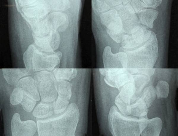
Fig. 9.1 Radiographs of the scaphoid. The radiographs show the lateral view, semiprone view, posteroanterior view, and semisupine view of the scaphoid. The semisupine view shows that the fracture is distal to the ridge and the semiprone view demonstrates the gap at the fracture site, indicating that this fracture is unstable.
If the fracture cannot be seen on any of these views but there is a high suspicion of one because of the nature of injury and local tenderness and pain on ulnar deviation of the wrist, then the surgeon may decide to perform a CT scan immediately. The CT scan identifies the location of the fracture and shows any displacement of the fracture. Such a displacement is usually seen on the dorsal surface on sagittal images
Transverse sections of the scaphoid obtained in a new scanner with 64 or 128 slices with a 50% overlap permit multiplanar reconstruction in the plane of the scaphoid. The CT scan is obtained with the patient placing the hand in a “Superman” position supported on a foam section. Sagittal-plane images are reconstructed. This allows assessment of bony architecture and the location and displacement of the fracture can be assessed as well (▶ Fig. 9.2).
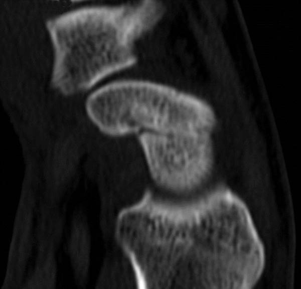
Fig. 9.2 Sagittal section of a CT scan obtained in the plane of the scaphoid helps define the fracture, which extends proximal to the scaphoid ridge with no obvious displacement. Such a fracture is very likely to heal even if immobilized for only 4 weeks in a below-elbow cast with the thumb left free, and the patient can be encouraged to return to most forms of work.
Classification The scaphoid fracture can be classified using one of three systems. No one classification is clearly predictive of union or helps determine treatment.
The Russe13 classification based on the inclination of the fracture line does not predict healing. Fractures may be horizontal oblique, transverse, or vertical-oblique. The vertical-oblique fractures account for only 5% of fractures. This pattern has the most shear force across the fracture site and may be unstable. Horizontal-oblique types have the most compressive force across the fracture site, and transverse fractures have both compressive and shear forces.
The Mayo Clinic classification divides scaphoid fractures on the basis of their location into proximal (30% of fractures), middle (65% of fractures), and distal (5% of fractures). Within the distal part, the classification further divides them into fractures that extend into the distal articular surface and those limited to the distal tubercle. Both these types will be seen well in the semiprone view. The location of the fracture influences both the union rate and time taken to heal. The rate of union in proximal, middle, and distal one-third scaphoid fractures is 64%, 80%, and 100%, respectively.14
The Herbert and Fisher classification15 is supposedly based on fracture stability. The type A Herbert classification fracture is a stable acute fracture, and a type B is an unstable acute fracture. However, stable fractures include fractures of the tubercle (A1) and an incomplete fracture of the waist (A2). These fractures can be treated nonoperatively. By this definition, all waist fractures of the scaphoid are unstable. All other types of fractures “may require surgical treatment.” Type B (acute unstable fractures) is further subdivided into B1 (oblique fractures of the distal one-third); B2 (displaced or mobile fractures of the waist); type B3 (proximal pole fractures); type B4 (fracture dislocations); and B5 (comminuted fractures).
Another study defined three main fracture patterns: those involving (1) the “surgical waist,” (2) the dorsal sulcus, or (3) the proximal pole. The fracture line extending to the dorsal sulcus was at 45° to the surgical waist and so was in the long axis of the bone. This group was further subdivided into three, with the fracture line passing proximal to, distal to, or on both sides of the apex. The butterfly fragment in the third subgroup usually showed displacement and comminution. These authors suggest that the fracture extending to the sulcus is much less stable than a surgical waist fracture and more likely to lead to the “humpback” flexed malunion.16
9.5 Management
Related posts:
 Proximal Row Fractures (Other Than Scaphoid and Pisiform Fractures): Triquetrum and Lunate Fractures
 Natural History of Traumatic TFCC Tears
 Role of Hand Therapy in the Treatment of Distal Radius Fractures
 Intra-articular Fractures of the Distal Radius (AO type C3), (Dry) Arthroscopic Approach
 Galeazzi Fracture-dislocation
 Unstable Ulnar Styloid Fracture
Stay updated, free articles. Join our Telegram channel

Full access? Get Clinical Tree


