This article reviews commonly performed injections about the foot and ankle region. Although not exhaustive in its description of available techniques, general approaches to these procedures are applicable to any injection about the foot and ankle. As much as possible, the procedures described are based on commonly used or published techniques. An in-depth knowledge of the regional anatomy and understanding of different approaches when performing ultrasonography-guided procedures allows clinicians to adapt to any clinical scenario.
Key points
- •
Ultrasound-guided injections about the foot and ankle can be used to assist in the alleviation of pain related to disorders of joints, bursae, tendons, and neurologic structures.
- •
Improved accuracy with ultrasound-guided injections about the foot and ankle allows these procedures to aid in the confirmation of a diagnosis as well as to improve safety, especially when performed adjacent to neurovascular structures.
- •
Clinicians should be familiar with alternative approaches for various ultrasound-guided procedures about the foot and ankle, because challenging individual patient anatomy or other factors may warrant modification of technique.
Tibiotalar (ankle) joint injection
Regional Anatomy
The tibiotalar joint is a synovial hinge joint formed by the articulation of the tibia and fibula with the underlying talus. The joint recess extends proximally from the inferior tibial margin by a mean of 19.2 mm. An intra-articular extrasynovial fat pad lies within the anterior recess. In addition, the ankle joint communicates with the posterior subtalar joint in 13.9% of cases. From medial to lateral, the tibialis anterior tendon, extensor hallucis longus tendon, dorsalis pedis artery and adjacent deep peroneal nerve, extensor digitorum longus tendon, superficial peroneal nerve, and peroneus tertius tendon overlie the anterior ankle joint.
Patient and Ultrasound Machine Positioning
- •
Patient: supine on table, injected side closest to provider
- •
Ultrasound machine: ipsilateral to involved side/provider
Transducer Type
- •
High-frequency linear-array: small footprint preferred
Needle Choice
- •
Injection only: needle 25 to 30 gauge, 25 to 38 mm
- •
Aspiration: at least 18-gauge, 38-mm needle
Injectate
Solution of local anesthetic plus corticosteroid (eg, 3 mL of 0.2% ropivacaine and 1 mL of 10 mg/mL triamcinolone).
Approaches
Long axis to joint in plane with transducer
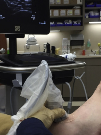
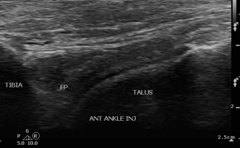
Transducer is aligned in a sagittal plane with the joint overlying the dome of talus ( Fig. 1 ). The needle is advanced in an anterior-to-posterior direction deep to the fat pad and superficial to the articular cartilage on the dome of talus, preferably avoiding overlying tendons and neurovascular structures ( Figs. 2–4 ).


Short axis to the joint in plane with transducer (the author’s preferred approach)
The transducer is aligned in an axial plane with the joint overlying the dome of talus. The needle is advanced in a lateral-to-medial direction deep to the fat pad and superficial to the articular cartilage on the dome of talus, preferably avoiding overlying tendons and neurovascular structures ( Figs. 5–7 ).
Long axis to the joint out of plane with transducer (technically more challenging)
The transducer is aligned in a sagittal plane with the joint overlying the dome of talus. Using a walk-down technique from superficial to deep, the needle is advanced in either a lateral-to-medial or an anterior-to-posterior direction deep to the fat pad and superficial to the articular cartilage on the dome of talus, preferably avoiding overlying tendons and neurovascular structures ( Figs. 8 and 9 ).
Tibiotalar (ankle) joint injection
Regional Anatomy
The tibiotalar joint is a synovial hinge joint formed by the articulation of the tibia and fibula with the underlying talus. The joint recess extends proximally from the inferior tibial margin by a mean of 19.2 mm. An intra-articular extrasynovial fat pad lies within the anterior recess. In addition, the ankle joint communicates with the posterior subtalar joint in 13.9% of cases. From medial to lateral, the tibialis anterior tendon, extensor hallucis longus tendon, dorsalis pedis artery and adjacent deep peroneal nerve, extensor digitorum longus tendon, superficial peroneal nerve, and peroneus tertius tendon overlie the anterior ankle joint.
Patient and Ultrasound Machine Positioning
- •
Patient: supine on table, injected side closest to provider
- •
Ultrasound machine: ipsilateral to involved side/provider
Transducer Type
- •
High-frequency linear-array: small footprint preferred
Needle Choice
- •
Injection only: needle 25 to 30 gauge, 25 to 38 mm
- •
Aspiration: at least 18-gauge, 38-mm needle
Injectate
Solution of local anesthetic plus corticosteroid (eg, 3 mL of 0.2% ropivacaine and 1 mL of 10 mg/mL triamcinolone).
Approaches
Long axis to joint in plane with transducer
Transducer is aligned in a sagittal plane with the joint overlying the dome of talus ( Fig. 1 ). The needle is advanced in an anterior-to-posterior direction deep to the fat pad and superficial to the articular cartilage on the dome of talus, preferably avoiding overlying tendons and neurovascular structures ( Figs. 2–4 ).
Short axis to the joint in plane with transducer (the author’s preferred approach)
The transducer is aligned in an axial plane with the joint overlying the dome of talus. The needle is advanced in a lateral-to-medial direction deep to the fat pad and superficial to the articular cartilage on the dome of talus, preferably avoiding overlying tendons and neurovascular structures ( Figs. 5–7 ).
Long axis to the joint out of plane with transducer (technically more challenging)
The transducer is aligned in a sagittal plane with the joint overlying the dome of talus. Using a walk-down technique from superficial to deep, the needle is advanced in either a lateral-to-medial or an anterior-to-posterior direction deep to the fat pad and superficial to the articular cartilage on the dome of talus, preferably avoiding overlying tendons and neurovascular structures ( Figs. 8 and 9 ).


