Fig. 2.1
(a) Hyperextension injury; (b) clinical presentation of acute turf toe
2.2 Etiology
A flexible shoe in combination with a hard artificial surface was considered the source of the turf toe injury in the 1970s.3,9,10 Even with the return to natural grass from artificial surfaces and third-generation artificial surfaces, this injury is still present. Risk factors for turf toe that have been investigated include: increased shoe sole to surface friction with a fixed forefoot, increased shoe flexibility, reduction in artificial turf’s ability to absorb force with wear.4,9 Valgus and varus stress mechanisms have also been theorized as causative factors of turf toe injuries.5 One possible risk factor that has not been investigated is chronic, repetitive, low-grade stress across the first MTPJ with physical activity of pushing, pulling, and jumping. These ballistic movements may contribute to inflammation of the first MTPJ causing a low-grade sprain on surfaces of varying rigidity. With repeated practices and games throughout a season including off-season workouts, overuse and breakdown of the joint tissue may occur. If 90% of the body’s weight is translated through the forefoot and 40–60% of the weight being placed across the great toe joint, overuse due to repetitive stretching or excessive tension on the plantar capsule ligaments could cause breakdown and disruption. An additional factor would include the increased physical ability of the modern athlete, being faster and stronger, intensifying the speed and velocity of ballistic movements.
2.3 Clinical Evaluation
Evaluation of the acute or subacute injured great toe joint needs to occur in a systematic, concise, and accurate manner. Initial exam may be thwarted by pain and splinting and thus, serial examination as acute symptoms resolve is mandatory. Each anatomical structure of the joint should be considered injured until proven otherwise. Identifying all structures that are potentially involved and determining the severity or lack thereof will allow the specialist to accurately grade the initial injury(s), outline an appropriate treatment program, determine a return to sports timeline, and to predict long-term morbidity. These goals are the cornerstone to the treatment of athletes.
The initial assessment includes ascertaining the mechanism and timing of the injury. Observe for any generalized deformity of the toe, the extent of joint effusion, the soft tissues around the distal first ray, and the possible presence and location of ecchymosis and swelling. Neurovascular compromise is not common except for extreme examples as seen with nonreduced dislocations. The author believes that ecchymosis is a hallmark clinical indicator of either a fracture or rupture of ligamentous or tendon structures. Ecchymosis plantarly increases the suspicion for sesamoid fracture and disruption of the plantar capsule-ligamentous structures or tear of the flexor hallucis brevis (FHB) muscle belly. Observation of ecchymosis dorsally may indicate dorsal tearing of the joint capsule, dorsal articular injury, or dorsal cortical fracture of the metatarsal. Palpation of the dorsal metatarsal head and neck along with the dorsal aspect of the base of the proximal phalanx provide suspicion for periarticular fracture. Further, palpation of the sesamoids and the long and short flexor and extensor tendons will provide information in regard to possible injury or fracture. Following palpation of the joint structures, manual manipulation of the joint should be performed. Specifically the medial and lateral collateral ligaments along with the phalangeal sesamoid ligaments provide controlled range of motion, proprioception and structural stability of the joint. Each ligament can and should be evaluated for the presence of pain and varying degrees of instability. A universal grading system should be applied to each ligament as increasing degree of injury will prolong return to sport and increase morbidity. A circumferential examination of the MTPJ should be performed beginning dorsal, medial then lateral and finally, plantar. The dorsal ligaments are assessed with plantar flexion of the joint, while the collaterals are evaluated with varus and valgus force with the hallux in a rectus position. Valgus stress can result in a ruptured medial joint capsule including the medial collateral ligament, abductor hallucis insertion and sesamoid apparatus (Fig. 2.2). The plantar ligaments are assessed by performing a Lachman-type maneuver. Again, while each ligament is evaluated (stressed), the presence or absence of pain as well as the amount of instability should be noted. Comparing stress evaluation of the noninjured side will at times be helpful. Tendonous injuries to the great toe will primarily involve the three main tendons of the great toe joint, the extensor hallusis longus (EHL), flexor hallusis longus (FHL), and FHB. Each tendon should be assessed for integrity and strength. In the authors’ experience, injury to tendons other than the FHB has not been encountered. As the exam is carried out, a clinical scorecard of the noninjured and injured structures and severity should be kept. The clinical exam will be prognostic for further imaging modalities.
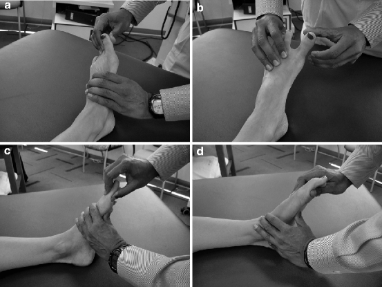
Fig. 2.2
(a) Abduction stress test to assess medial collateral ligament/medial capsule/abductor hallucis; (b) Adduction stress test to assess lateral collateral ligament/lateral capsule/adductor hallucis; (c) “Lachman” of first MPJ to assess plantar plate; (d) FHB strength/integrity assessment
2.4 Imaging of Turf Toe Injuries
When imaging turf toe injuries, a protocol is necessary for accurate diagnosis. These radiographs should be evaluated for overt or small avulsion fractures of the distal first ray as well as mal-alignment, chondral injury, and sesamoid position. Radiographs of the contralateral foot should be performed for comparison. Initial imaging of the injured foot begins with plain radiographs taken in a standard anteroposterior (AP) projection, lateral oblique projection, and lateral views of the foot in weight bearing. Sesamoid position on an AP radiograph can provide information regarding the presence of sesamoid injury such as dislocation, retraction of the sesamoids (Fig. 2.3), disruption of the inter-sesamoid ligament, or fracture of the sesamoids. An additional image is the axial sesamoid view, with the foot weight bearing, the heel raised with the toe dorsiflexed and the radiographic beam being introduced from posterior 90° to the foot.
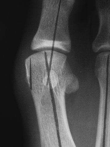
Fig. 2.3
X-ray of dislocation with sesamoid retraction
Additional radiographs can be taken performing provocative maneuvers to the first MTPJ, stressing the plantar joint capsule and surrounding ligamentous attachments. A Lachman’s type maneuver or dorsiflexion stress maneuver can be performed at the joint to accentuate instability. Performing dorsiflexion stress maneuvers can reveal retraction of the sesamoids or disruption of the plantar plate on the metatarsal showing evidence of metatarsal sesamoid ligament disruption. In addition, fluoroscopic imaging can be used to evaluate range of motion of the first MTPJ and demonstrate instability.
There is very little information defining the indications of magnetic resonance imaging (MRI) for turf toe injuries. But clearly, this advanced imaging modality will allow the clinician to appreciate the full spectrum of the injury and serve as an adjunct to the clinical exam. MRI will allow for each potentially injured structure to be evaluated for the presence or absence of injury and severity (Fig. 2.4). MRI imaging of the MTPJ complex using a non-fat-suppressed T1-weighted or proton density–weighted sequence in three standard planes is recommended.6 In addition, according to Crain et al.,6 a proton density–weighted fat-suppressed or short tau inversion recovery (STIR) sequence in all three planes is recommended for optimal evaluation of the turf toe pathology. A STIR sequence should be used to evaluate acute pathology. Proton density–weighted fat-suppressed images provide better resolution than STIR with shorter imaging time and also provide more detail of the anatomy than heavily fat-suppressed T2-weighted images and STIR sequences. To optimize the resolution with lower field MRI scanners, that field of view should range from 13 to 15 cm with a slice thickness of no less than 3.5 mm of STIR images.6 Higher field MRI scanners such as 3.0 T magnet provide better definition and detail. Injury of the metatarsal sesamoid ligament and phalangeal sesamoid ligament is best visualized on sagittal slices. The inner sesamoid ligamentous anatomy is best visualized on axial images. The most common injured soft tissue structures along the plantar first MTPJ capsule are the phalangeal sesamoid ligamentous attachments. In MRI, evaluation of the injured plantar phalangeal sesamoid ligamentous structures will reveal incontinuity or edema. Once the clinical exam is correlated with radiographic and possible advanced imaging, the clinician can now truly understand the full spectrum of the injury.
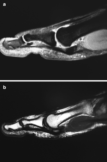
Fig. 2.4
(a) MRI showing plantar first MTPJ plate rupture without sesamoid fracture; (b) Repaired plantar plate
2.5 Classification of Turf Toe
Turf toe injuries are classified on the basis of severity. To date, there is no grading system that is truly comprehensive and takes into consideration injuries to structures other than the ligaments. As can be appreciated, if there is injury to osseous, articular, or tendonous structures, each will have its own spectrum of injury, treatment protocol, and morbidity. A general assumption can be made that with increasing degrees and involvement of ligamentous structures, there is a greater chance of associated injuries to occur. It is often the lack of appreciation of these associated injuries that will lead to an inaccurate initial diagnosis and prognosis. In treating athletes, the specialist is tasked with working with a small margin of error. This margin will decrease as the level of the athlete increases. Establishing the grade of turf toe injury from clinical evaluation and imaging protocol will provide the most accurate initial diagnosis and treatment, shorten the convalescent period, reduce the time to return to play, and predict possible morbidity that may affect long-term performance.
Related posts:


Stay updated, free articles. Join our Telegram channel

Full access? Get Clinical Tree


