Treatment of Tarsal Coalitions
Patient Selection

Figure 1Clinical photographs show a patient with a unilateral calcaneonavicular coalition. In the supine position, the affected foot (arrows) fails to form an arch (A) and has diminished plantar flexion (B).
Patients present with myriad symptoms: foot pain, foot deformity, recent or prior foot or ankle injuries
Suspect tarsal coalition in teenager with multiple ankle sprains
Careful history, physical examination help identify coalitions
Stiff flatfoot is hallmark of tarsal coalition; can be dramatic when unilateral (Figure 1, A)
Calcaneonavicular coalitions
Restricted subtalar motion
Palpable bony ridge in sinus tarsi
Restricted plantar flexion (Figure 1, B)
Talocalcaneal coalitions
Restricted subtalar motion
Tender bony prominence around sustentaculum tali
Author asserts nonsurgical management has no long-term benefit; recommends excision for all young patients; temporary cast relief outweighed by altered biomechanics, long-term effects, including adjacent joint degeneration
Preoperative Imaging

Figure 2Radiographs demonstrate typical findings of a tarsal coalition. A, Internal rotation oblique view demonstrates the calcaneonavicular coalition (arrow). B, Lateral view shows the C-sign, which indicates a talocalcaneal coalition. This sign is present when the posterior margin of the talus appears to be continuous with the sustentaculum tali (arrows). The C-sign also can be seen in flexible flatfoot.
Coalition types have classic radiologic signs: anteater sign for calcaneonavicular coalitions (Figure 2, A), C-sign for talocalcaneal coalitions (Figure 2, B)
CT of both feet preferred for all preoperative cases; CT with three-dimensional reconstructions even more helpful
MRI can identify fibrous coalitions
Procedure
Room Setup/Patient Positioning
Position foot near end of table so team can be seated
Place sterile tourniquet; perform Esmarch exsanguination
Special Instruments/Equipment/Implants
C-arm
Kerrison rongeurs (3 or 4 mm)
Osteotomes
High-speed burr (3 or 4 mm)
Surgical Technique
Calcaneonavicular Coalition Resection
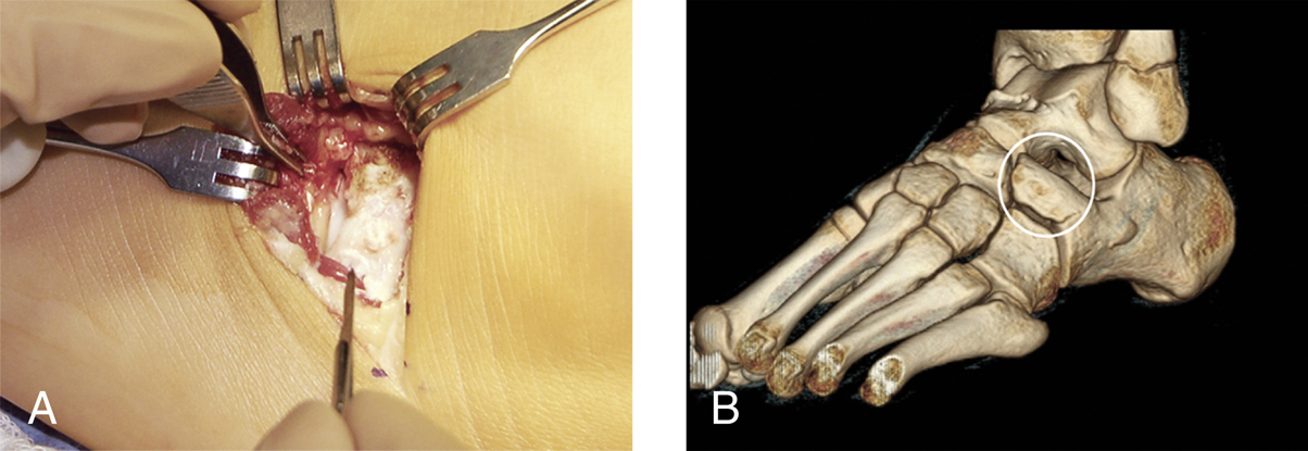
Figure 3Images demonstrate the exposure for calcaneonavicular coalition resection. A, Intraoperative photograph shows the extensor digitorum brevis elevated off the coalition by proximal release at its origin. The coalition is visible directly beneath the elevated muscle. B, Three-dimensional CT scan is used to ensure that the intended plane of dissection is the correct one.
Stay updated, free articles. Join our Telegram channel

Full access? Get Clinical Tree


