Arthroscopic Treatment of Anterior Ankle Impingement
Keywords
• Ankle arthroscopy • Anterolateral ankle impingement • Anterior ankle impingement • Chronic ankle pain • Soft-tissue ankle impingement
Ankle impingement was first described in 1942 by Morris as athlete’s ankle and was later coined footballer’s ankle by McMurray.1,2 Today, anterior ankle impingement syndrome describes the condition in which anatomic structures become entrapped in and around the ankle joint leading to chronic pain and potentially decreased range of motion. Ankle impingement can be caused by either soft-tissue or osseous pathology and is often classified by the location within the ankle joint. The most common areas of ankle impingement include anterolateral, anterior, and posterior. This article discusses common etiologies, clinical and diagnostic evaluation, treatment options, and outcomes for anterior ankle impingement.
Anterolateral ankle impingement
There are more than 23,000 inversion ankle sprains daily in the United States and it is the most frequently observed injury in athletics.3 Approximately 20% of patients will have residual or chronic ankle pain.3 Studies have shown that 50% of all basketball players with ankle sprains had residual symptoms, with 15% experiencing compromised playing performance.4–6 It is estimated that 3% of all ankle sprains lead to anterolateral impingement.7 Some think the term chronic ankle sprain should be replaced by anterolateral impingement of the ankle.8 The 3 most common types of soft-tissue lesions that have been documented to cause anterolateral impingement are meniscoid lesions, synovitis, and distal anterior inferior tibiofibular ligament impingement.
Meniscoid Lesion
In 1950, Wolin and colleagues9 were the first to describe a cause for anterolateral impingement. The study consisted of 9 patients with chronic pain and swelling to the anterolateral aspect of the ankle after an inversion ankle sprain. Upon surgical examination, patients were found to have a soft-tissue mass consisting of hyalinized tissue that the investigators identified as meniscoid lesions because of its resemblance to a torn knee meniscus. These fibrous adhesions are well-developed bands of scar tissue that occur from the anterior talofibular ligament and extend into the ankle joint, resting in the lateral gutter (Fig. 1). Upon removal of the meniscoid lesions, Wolin noted improvement of the patient’s symptoms. Initially thought to be torn ends of a ligament, histopathologic analysis revealed no ligamentous tissue.8 Several investigators have noted and attributed meniscoid lesions to the cause of anterolateral ankle pain.10,11
Synovitis
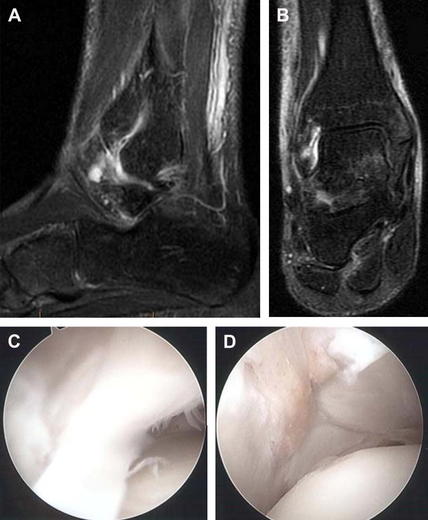
Synovitis may also occur after an inversion ankle sprain.8 The sprain may damage the anterior talofibular ligament (ATFL), anterior inferior tibiofibular ligament (AITFL), or the calcaneal fibular ligament without causing frank instability. Improper treatment and rehabilitation will lead to inadequate healing, allowing repetitive motion to cause inflammation at the ligament ends. Over time, continued activity allows the synovium to enlarge, causing impingement in the lateral gutter and chronic lateral ankle pain (Fig. 2).8 Hemorrhagic synovitis can occur because of mild capsule tearing resulting in hematoma formation. The hematoma eventually is resorbed by the synovium, causing synovitis. In Ferkel and Fisher’s12 study of 31 patients with anterolateral ankle impingement, all had either hypertrophic or hemorrhagic synovitis. Subsequent studies have noted a majority of patients having synovitis present with anterolateral ankle impingement. Recently, an intraoperative classification was developed to describe impingement in the lateral gutter. This classification was based on the degree of obstruction in the lateral gutter. It ranged from normal (no abnormal soft tissue present) to severe (excessive anterolateral soft tissue preventing any visualization before shaving/debridement). In a study consisting of 41 patients with anterolateral impingement, 7 patients were noted to have what the investigators termed “synovial shelves,” which was described as a hypertrophic band of synovium extending from the anterolateral aspect of the fibula over the lateral shoulder of the talus to the anterior ankle.13 It has been noted that early resection of impinging synovium inhibits the progression of the cascade to chronic synovitis and scar-tissue formation.14
Distal Fascicle of Anterior Inferior Tibiofibular Ligament (Bassett’s Ligament)
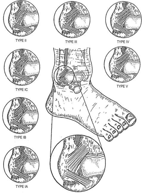
Bassett and colleagues15 discovered another cause of impingement after inversion ankle sprains when they described the distal fascicle of the AITFL. Nikoloponlous16 originally described Bassett’s ligament in 1982 as an accessory AITFL because of the distinct fibrofatty septum between the two ligaments. The research, however, was never published in the English literature.16 The incidence of the distal fascicle of the AITFL ranges from 21% to 92%, with variation most likely caused by varying descriptions of the distal fascicle.15–19 One investigator noted that it is most likely a common finding and only becomes pathologic after ankle mechanics become changed. The distal fascicle is found inferior and parallel to the AITFL, with its length ranging from 17 to 22 mm, a thickness of 1 to 2 mm, and width of 3 to 5 mm. An anatomic report by Ray and Kriz19 devised a classification system describing 5 distinct types (Box 1, Fig. 3).
Box 1 AITFL classification system
| Type 1 | There are multiple fascicles (more than 3) with or without small gaps between adjacent fascicles. A1: the inferior fascicle is separated from the rest of the ligament by a gap and possesses its own distinct proximal and distal attachments. B1: the inferior fascicle is not completely separated from the rest of the ligament by a gap. Either its proximal or its distal attachment is continuous with the rest of the ligament. |
| Type 2 | There are 3 fascicles or less. There is a distinct inferior fascicle with both its proximal and distal attachments separate from the rest of the ligament. The inferior fascicle is separated completely from the main portion of the ligament by a gap. |
| Type 3 | There are 3 fascicles or less. There is a distinct inferior fascicle with either its proximal or its distal attachment continuous with the rest of the ligament. A gap does not completely separate the inferior fascicle from the rest of the ligament. |
| Type 4 | There are 3 fascicles or less. The lower portion of the ligament possesses an inferior fascicle with both its proximal and distal attachments for the rest of the ligament. |
| Type 5 | There are 3 fascicles or less. There is a ligament with no separations or gaps within its structure. It may or may not possess a fascicular arrangement. |
Adapted from Ray RG, Kriz BM. Anterior inferior tibiofibular ligament. Variations and relationship to the talus. J Am Podiatr Med Assoc 1991;81(9):479–85; with permission.
The distal fascicle runs in intimate contact with the anterolateral corner of the talus. Ray and Kriz noted lateral border impingement of the talar dome in 35 of 46 specimens. In anatomic studies, associated cartilage damage is noted between 17% and 89% and has been confirmed during arthroscopic treatment.15–19 Bassett attributed the cartilage damage as a result of increased ankle laxity from an inversion ankle sprain.15 The attenuation of the ATFL allows the anterior lateral aspect of the talus to extrude anteriorly during dorsiflexion, causing fraying of the cartilage. Akseki and colleagues18 transected the ATFL in cadavers and placed the ankle through range-of-motion testing. This process led to 4 differences where distal fascicles were already in contact with the talus in a neutral position (42 of 47 specimens).
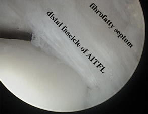
The variation of width, length, and obliquity may also be related to pathology. Akseki determined that the mean width and length were significantly higher in fascicles that bent during dorsiflexion. It was also noted that the more distal the insertion of the fascicle near the ATFL, the more often impingement occurred.19 Bassett noted that bending of the fascicle on dorsiflexion greater than 12° might be the source of thickening and impingement. It has been noted that manual distraction of the ankle joint relieves contact and impingement of the fascicle on the talus.15 Therefore, the investigators concluded that the use of intraoperative distraction might cause the surgeon to miss a pathologic distal fascicle (Fig. 4).
Anterior ankle impingement
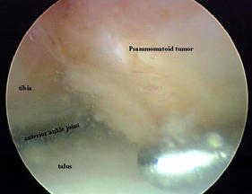
Anterior ankle impingement is most commonly caused by an osseous etiology presenting as pain in the anterior medial aspect of the ankle. Although there are small case studies of soft tissue and space occupying lesions causing anterior ankle impingement (Fig. 5), this section focuses on osseous pathology.20,21
Osseous
Formation of tibiotalar osteophytes at the anterior aspect of the ankle is a common cause of chronic ankle pain.22 Anterior ankle spurring is a common occurrence in the athletic community, ranging from 45% to 60%.23 Historically, 2 hypotheses for the etiology of anterior osteophyte formation have been described. According to McMurray,2 anterior osteophyte formation develops from repeated capsular traction while having the foot in a maximally plantar-flexed position. The investigator used the example of an athlete kicking a soccer ball. Other investigators have supported this theory. However, recent anatomic studies have demonstrated this hypothesis to be unfeasible.24
One cadaveric study measured the width of the non–weight-bearing tibial cartilage rim, and the distance from the tibial and talar cartilage from capsular structures were measured. The results revealed that the tibial cartilage to capsule distance was 4.3 mm and talar cartilage to capsule was 2.4 mm. The width of the tibial non–weight-bearing cartilage was 2.4 mm. This study refutes the theory that osteophyte formation is caused by traction.24 Arthroscopically, tibial osteophytes are noted to be within the joint, and it is not necessary to reflect the capsule to remove the osteophyte. The cadaveric specimens were also manually manipulated to 15° dorsiflexion to study the soft-tissue components at the anterior joint space. Histopathologic analysis was performed and revealed a soft-tissue layer bordered by synovial cells forming a synovial membrane. The investigators concluded that anteriorly located soft tissue impinges between the osteophytes. This soft tissue becomes inflamed and is the source of ankle pain and not the osteophytes themselves.
The second hypothesis described by O’Donoghue,25 states that the osteophytes were caused by direct trauma during forcible dorsiflexion and not at extreme plantar flexion causing traction as initially stated by McMurray. The author noted patients to have pain on end range of dorsiflexion or as the author coined “drive.”25 Stoller and colleagues23 theorized that repeated impingement causes subperiosteal hemorrhages and subsequent bone growth.
It has been noted that the non–weight-bearing anterior cartilage rim undergoes osteophytic transformation.26 The investigator thought the osteophyte formation was caused by direct damage and recurrent microtrauma to the anterior cartilage rim. The same investigators performed a biomechanical analysis to determine if whether kicking a soccer ball could cause osteophyte formation by hyperplantar flexion or by direct impact.22 Results revealed that in 39% of kicking movements, maximum plantar flexion exceeded the subject’s static maximum plantar flexion. The soccer ball impact was noted at the anterior part of the medial malleolus in 76% of ball strikes, with ball velocity averaging 24.6 m/s and contact force of 1025 N. The investigators concluded that the impact location and force, not extreme plantar flexion, support the hypothesis that spur formation occurs from direct repetitive trauma to the anterior cartilage rim. This microtrauma may be compounded in patients with a history of supination injuries. A history of supination injuries has also been documented in 76% of patients with osteophyte formation in one study, and in another study the incidence of spurs was 3 times higher in an unstable ankle joint compared with a group without ankle instability.27
Clinical presentation
Physical examination reveals pain on palpation along the anterior lateral aspect of the talus, the lateral gutter, and the most inferior aspect of the tibiofibular articulation. Soft-tissue swelling is present in the anterolateral shoulder of the ankle joint and palpable masses are occasionally noted within the lateral gutter.9 Pain can be elicited with passive dorsiflexion and eversion. An audible click may also be heard during ankle range of motion. This click has been attributed to AITFL impingement. The “ankle impingement sign,” described by Malloy, uses thumb pressure over the lateral gutter while taking the ankle from a plantar-flexed position to maximal dorsiflexion. The investigators noted that if hypertrophic synovitis is present, this maneuver impinges the synovium between the neck of the talus and the distal tibia. This test has been reported to be 94.8% sensitive and 88.0% specific for synovial hypertrophy.28
Liu and colleagues29 noted 94% sensitivity and 75% specificity predicting anterolateral ankle impingement using their clinical examination. Impingement was considered positive when at least 5 of 6 clinical parameters were present:
The clinician must rule out other causes of anterior lateral ankle pain, such as osteochondral lesions of the talus, peroneal tendonitis or subluxation, tarsal coalitions, and subtalar joint dysfunction.30
In anterior ankle impingement, patients will describe pain on dorsiflexion with a subjective feeling of stopping or blocked dorsiflexion. McMurray2 observed patients having pain and difficulty kicking a soccer ball correctly at the medial aspect of the ankle. Pain was not elicited when the ball was kicked improperly with the toes. Ballet dancers will describe pain in demi-plié or grand-plié and with landing from a leap.31 On physical examination, pain on palpation is noted to the medial aspect of the ankle joint. With the foot in maximum plantar flexion, osteophytes may be palpable medial to the tibialis anterior tendon or along the anterior rim of the tibia because of the thin subcutaneous tissue.26
Radiographic Evaluation
In patients with anterolateral impingement, plain radiographs should be performed to rule out fractures, widening of the ankle mortise, or arthritic changes. Stress radiographs can be used to rule out ligament laxity. In patients with suspected anterior ankle impingement, accurate detection and localization of spurs are necessary for preoperative planning.26 Standard anteroposterior and lateral views should be performed to detect anterior tibial and talar spurring. Scranton and McDermott32 devised a classification for spur formation: stage I, osteophyte less than 3 mm; stage II, osteophyte greater than 3 mm; stage III, anterior tibial osteophyte with talar osteophyte (kissing lesion); and stage IV, panarthritis. Standard lateral films have been shown to be capable of detecting only 40% of tibial and 32% of talar osteophytes. The anteromedial notch and anteromedial contour can inhibit the detection of anteromedial osteophytes.33 Tol and colleagues33 devised an oblique radiograph (45/30 anteromedial impingement [AMI] view) where the beam is tilted 45° in the craniocaudal direction with the leg in 30° of external rotation and the foot plantar flexed. In a study of 60 patients with anterior impingement, lateral radiographs were only able to detect lateral spurring in 58% of ankles. When detecting both tibial and talar osteophytes, the lateral view was 40% and 32% sensitive. When combined with the 45/30 AMI view, sensitivity increased to 85% and 73%.
Stay updated, free articles. Join our Telegram channel

Full access? Get Clinical Tree


