Arthroscopic Treatment of Ankle Osteochondral Lesions
Keywords
• Osteochondral lesion • Talar dome lesion • Ankle • Arthroscopy
Osteochondral lesions (OCLs) of the ankle represent a host of pathologies, from subtle chondromalacia to full-thickness defects with underlying cystic changes and osteonecrosis. Frequently these lesions are traumatic in origin, most commonly occurring after an acute ankle sprain; however, atraumatic mechanisms have been described. Osteochondral lesions of the talus (OLT) are more common than lesions of the tibial plafond. In their landmark paper, Berndt and Harty1 delineated both a classification system and a clarification of the behavior of these injuries, focusing on mechanism and location of the lesion.
Pathophysiology
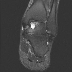
The pathophysiology of OCLs must be appreciated to fully understand why the various treatment modalities are effective and when to use them. The initial insult involves some level of joint or articular damage, whether from trauma or other metabolic, genetic, vascular, or idiopathic processes.2 Many lesions are often traced back to a specific ankle sprain, ankle fracture, or other lower extremity trauma.3 Alternatively, nonspecific repetitive microtrauma may generate an OCL over time, or asymptomatic necrotic lesions may become symptomatic with subtle injuries. Regardless of the inciting event or baseline pathology, the processes through which these lesions become symptomatic are the same. Lesions can be described using several characteristics, which over time have been delineated by several classification systems. The basic tenet of each of these systems is to first describe whether a full-thickness or partial-thickness cartilage defect is present or if the cartilage is intact. The quality and condition of the subchondral bone plate and the underlying trabecular bone are important to know. A fragment of bone may be attached to the disrupted cartilage. The subchondral plate may be fractured or compacted and the underlying bone may have become sclerotic. Subchondral cyst formation may have occurred. These features should be noted and may offer clues as to the physiologic process and appropriate treatment (Fig. 1). Whether the fragment is partially or fully detached or displaced should also be noted. Understanding these dynamics of the lesion provides clues to the origin and may assist in directing treatment.
Partial-thickness or full-thickness flaps of cartilage that have separated from the underlying subchondral bone are created through shearing forces and are not amenable to being left alone to repair themselves because of lack of blood supply. They will act as an irritant in the joint space, promoting synovial inflammation and subsequent symptoms. Sometimes this synovitis is more symptomatic to the patient than the lesion itself. These cartilage flaps have been recently called chondral-separated lesions, in contradistinction to osteochondral-separated lesions.4 This latter type of lesion is more commonly referred to as an osteochondral fracture and may have a better chance of forming fibrocartilage because of its retained blood supply from the subchondral bone.
Cysts may form with either chondral or osteochondral lesions when the subchondral plate is compromised. Where small defects in the subchondral plate exist, repetitive loading from normal weight-bearing activates forces the synovial fluid under high pressure into the subchondral bone, which over time creates a cyst.5,6 Cystic lesions may also be seen with apparently intact cartilage. This finding can be explained by a similar mechanism in which the subchondral plate is fractured and the fluid content of the cartilage is exsanguinated and forced into the subchondral bone with repetitive weight-bearing pressures. As the cyst develops and the integrity of the subchondral plate collapses, the overlying cartilage becomes soft because of the absence of this supportive structure. Over time, as these cavities are continually filled with fluid under pressure, the bone reabsorbs, creating a subchondral cyst, which may become sclerotic as the exposed bone remodels.5,6 Whether these lesions are caused by trauma or local necrosis, they may evolve to include sclerotic areas of bone with associated subchondral cyst formation. The theory of these nuances led to the development of many of the operative treatments currently used. Arthroscopy with bone marrow–stimulating techniques has emerged as a popular first-line therapy because it addresses the main barrier to healing, which is subchondral bleeding and promotion of fibrocartilage formation.
A basic knowledge of cartilage anatomy and physiology helps in understanding of the goals, mechanism, and limitations of arthroscopic treatment of OCLs. Native articular cartilage consists of hyaline cartilage. Hyaline cartilage is unique in that its matrix consists of primarily type II collagen, which has improved tensile strength over type I collagen, the predominant component of fibrocartilage. Hyaline cartilage, however, cannot be regenerated once injured. Fibrocartilage is the natural repair and physiologic alternative. Hyaline cartilage has abundant water content, accounting for approximately 75% of the cartilage matrix.5,6 The matrix also contains fillers such as proteoglycans that aid in resisting compressive forces. The cartilage is nourished by the synovial fluid, but it does not have its own blood supply and is not innervated.5,6 Articular cartilage can be divided into four zones.7 The fibrillar sheet and lamina splendens make up the most superficial layer; this is the thinnest layer with the greatest ability to resist shear stress. Once violated, degradation and fibrillation become progressive, manifesting as a combination of any of the lesions previously described, depending on local physiology and external stress. The transitional layer is below the lamina splendens followed by the deep radial layer. The deep radial layer is the largest layer distributing force and resisting compression. The deepest layer is the calcified cartilage, the beginning of which is called the tidemark, which separates the hyaline cartilage from the underlying subchondral bone. This layer is significant in osteochondral repair procedures involving allograft or autograft material, because the tidemark level differs between different areas of individual joints and different joints themselves, thus having significant implications on loading and healing characteristics.
Diagnosis and Workup
Patients presenting with ankle OCLs may have a history of trauma and will describe vague symptoms such as swelling, deep ankle pain, instability, locking, or catching. The pain is typically difficult to reproduce on examination but can be confirmed with a response to a diagnostic ankle block. Lesions may be identified on plain radiographs. Ancillary imaging studies are useful when a high clinical suspicion exists or further clarification of the extent and nature of the lesion is needed. These studies often assist in preoperative planning. Several imaging specific classification systems have been developed with this goal in mind. Bernt and Harty’s1 classification system is based on plain radiographs and includes four stages from compression of the cartilage (stage 1) through a displaced lesion (stage IV). Although this system is useful, it has little prognostic value and as many as 50% of OCLs are missed on plain radiographs, necessitating advanced imaging.8
CT, although it accurately assesses the extent of bone involvement, is unable to assess the extent of the chondral injury, which is important in preoperative planning. Ferkel and colleagues9 developed a classification scheme based on CT describing the osseous component with respect to cystic changes and communication with the joint surface.
MRI has gained popularity in its ability to delineate both the cartilage and bone extent of the lesion in addition to associated soft tissue pathology. Several MRI classification systems have been proposed, most of which stage lesions from chondral bruising through a detached fragment with a focus on the quality of the cartilage and the nature or absence of its attachments.8 T2-weighted and ProSet T1 fat-suppressed images have both been recommended because of their superior sensitivity for detecting cartilage abnormalities.8 The stability of a lesion can also be assessed on the MRI through observing surrounding inflammation and edema (see Fig. 1), although this is of unknown importance for preoperative planning and prognosis.
On T2-weighted images, increased signal intensity can be seen surrounding completely detached lesions, and bone edema may be present. These findings have been considered evidence of instability, which has been used as an operative indication; however, no clear correlation exists. In their recent work exploring why only some osteochondral defects in the ankle are painful, van Dijk and colleagues5 attribute painful lesions to the repetitive increased fluid pressures. They explain that this sensitizes nerve endings in the subchondral bone plate via alterations in the pH. MRI is the best imaging modality to detect evidence of high fluid pressures surrounding lesions, which manifest as high signal intensity around the lesion and bone marrow edema on fat-suppressed images. Therefore, if painful lesions are assumed to be painful because of instability, these MRI findings are consistent with both.
Plain radiographs, CT, and MRI are all intended to help with treatment selection and preoperative planning where indicated; however, MRI seems to offer the most useful information and should be performed in most cases. Surgeons are cautioned that MRI may exaggerate the extent of osseous involvement in OCLs.8 A threshold beyond which arthroscopy is unlikely to yield satisfactory results has been shown to exist around lesions greater than 1.5 cm2.10–12
When arthroscopy is used, arthroscopic-specific classification systems can be used and have been shown to have prognostic value.13 Several arthroscopic staging systems have been introduced. Pritsch14 introduced a three-stage system in 1986 describing the cartilage as intact, soft, or frayed. In 1995, Ferkel and colleagues13 introduced a more elaborate system that included stages A through F, in which A through C describe worsening grades of cartilage wear and stages D through F describe progressive lifting, detachment, and displacement of the fragment (Box 1).


