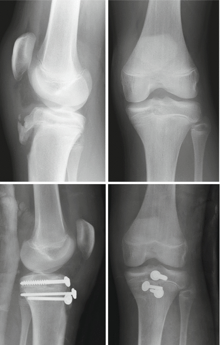
Figure 7.1
Radiographs of a distal femoral physeal injury and fixation using two parallel screws
A long leg cast should be applied for 4–6 weeks.
7.1.1.6 Follow-Up
K-wire pin sites should be checked at 1–2 weeks and the plaster cast replaced with a lightweight fibreglass cast.
K-wires should be removed at 3–4 weeks, especially trans-epiphyseal crossed K-wires as they may be intra-articular and predispose to septic arthritis.
All physeal fractures should be followed up for at least 1 year to look for a growth arrest.
7.1.1.7 Complications
These may be categorised as below:
Immediate:
Neurovascular injury.
Malreduction.
Compartment syndrome.
Early:
Re-displacement.
Infection.
Late:
Growth arrest or angular deformity.
Limb length discrepancy.
7.1.1.8 Synopsis
These are uncommon injuries (2–5% of physeal fractures) that need to be considered with a high index of suspicion. They should be treated with anatomical reduction and internal fixation to minimise late angular deformity or growth arrest.
7.1.2 Proximal Tibial Physeal Injuries
7.1.2.1 Introduction
Proximal tibial physeal injuries are rare due to intrinsic stability as a result of the overlapping fibula, the anatomy of the tibial tubercle and the metaphyseal attachment of the medial collateral ligament (MCL). Considerable forces are required to separate the proximal tibial physis and therefore concomitant injuries are often seen. Vascular compromise is common due to the popliteal vessels being tethered in this region, and so compartment syndrome is an important consideration.
The proximal tibial physis contributes 60% toward the growth of the tibia, amounting to approximately 0.9 cm per year. Unlike distal femur fractures, proximal tibial physeal injuries rarely succumb to growth arrest after injury.
7.1.2.2 Fracture Classification
The most common type of injury is a SH II, but towards end of growth the proximal tibial physis fuses from posterior to anterior, which may result in more SH III injuries.
7.1.2.3 Diagnosis
Patients present with a history of a direct force (such as following an RTA) or a hyperextension type injury. Examination reveals marked tenderness and swelling of the proximal tibia with a possible concavity at the level of the tibial tuberosity, if the metaphysis has been displaced posteriorly. As with all such knee injuries, always assess and document the neurovascular status, and observe the patient for a potential impending compartment syndrome.
7.1.2.4 Imaging
Begin with an AP and lateral of the knee, augmented with oblique films if the aforementioned views appear normal. Stress radiographs or CT/MRI may be helpful in diagnosis.
7.1.2.5 Treatment
Non-operative Treatment
Undisplaced fractures may be treated in an above-knee backslab, then converted to a fibreglass cast with slight knee flexion for 6 weeks total.
Always watch for compartment syndrome in the first few days.
Indications for Surgery
Anatomical reduction is necessary for the best result. Anything short should be considered for surgical intervention.
Even minimal posterior displacement of the metaphysis can occlude the popliteal artery, so thought must be given to treating posteriorly displaced fragments.
Displaced SH I and II fractures can be treated with closed reduction, followed by trans-epiphyseal crossed K-wires passed from the metaphysis into the epiphysis. Whenever any physis is crossed, a single pass technique is advocated. The wires should not cross at the fracture site. Open reduction may be required if the pes anserinus prevents closed reduction.
Displaced SH III and IV fractures will often require opening to ensure anatomical reduction and placement of K-wires or screws horizontally into the epiphysis, and proximal and parallel to the physis.
Fasciotomies are required if vascular compromise and compartment syndrome are identified.
7.1.2.6 Follow-Up
Backslabs should be removed and the wounds checked at 1–2 weeks. Full fibreglass casts should then be applied until the K-wires are removed at 3–4 weeks. The fibreglass above knee cast is discontinued at 6 weeks. Patients should be followed up for at least 1 year to observe for growth disturbance.
7.1.2.7 Complications
Immediate:
Compartment syndrome.
Neurovascular compromise.
Early:
Infection.
Malreduction.
Late:
Angular deformity.
Growth arrest.
Limb length discrepancy.
7.1.2.8 Synopsis
A rare physeal injury but often associated with other injuries and vascular compromise or compartment syndrome. Anatomical reduction and internal fixation is required to prevent complication.
7.1.3 Tibial Tubercle Avulsion
7.1.3.1 Introduction
Avulsion of the tibial tubercle apophysis represents a SH III injury. It is most commonly seen in boys aged 12–16 years and associated with jumping sports due to sudden eccentric loading of the extensor mechanism. A large periosteal sleeve may be avulsed with the tubercle. Tibial tubercle avulsion should not be confused with Osgood-Schlatter disease which is a chronic condition of repeated micro-avulsion of the superficial tuberosity, not involving the apophysis.
7.1.3.2 Classification
The most widely used classification system was devised by Ogden. The system takes into account the two ossification centres in the proximal tibia: the primary ossification centre (proximal tibial physis) and the secondary ossification centre (tibial tubercle physis) (Fig. 7.2).

Type I – fracture of the secondary ossification centre.
Types II – fracutre propagates to the junction with the primary ossification centre.
Type III – fracture crosses the primary ossification centre.
Modification A: undisplaced.
Modification B: displaced.
Figure 7.2
The Ogden classification
7.1.3.3 Diagnosis
Patients typically present following a jump or fall, followed by pain in the anterior aspect of their knee and inability to straight leg raise. Examination findings include swelling and tenderness over the tibial tubercle with the knee held in slight flexion. Some active knee extension may be present if retinacular fibres are intact. Patella alta may be noticed and the mobile bone fragment may be felt if it is large enough. Compartment syndrome has been reported.
7.1.3.4 Imaging
Lateral knee radiographs will confirm the injury and show the amount of displacement. Look for patella alta and if necessary, compare to the non-injured side.
7.1.3.5 Treatment
Type IA fractures (undisplaced) can be treated in a cylinder cast in full extension for 6 weeks.
Type 1B, 2 and 3 fractures should be treated by open reduction and internal fixation. Cancellous or cortical screws are placed through the tuberosity fragment horizontally across the metaphysis. Large periosteal sleeves can be reattached using suture anchors if desired (Fig. 7.3).