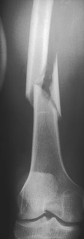
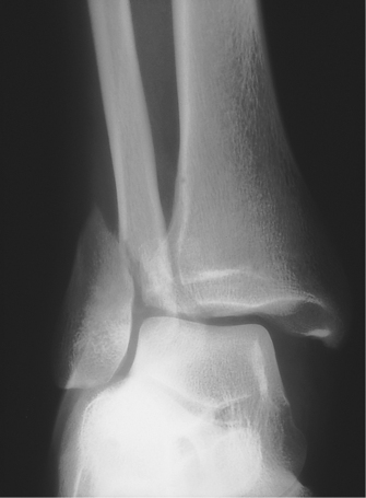
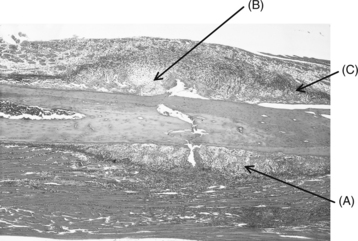
TRAUMA AND THE MUSCULOSKELETAL SYSTEM

10


TRAUMA AND THE MUSCULOSKELETAL SYSTEM


Andrew Ellis and Thomas Taylor


Introduction


Musculoskeletal injury frequently occurs in the setting of major trauma. The energy required to produce fractures, especially of diaphysial bone in young people, is considerable. Such energy is often associated with falls from a height, motor vehicle accidents, workplace accident or assault. Blunt trauma is more common but penetrating trauma (from bullets in particular) is not uncommon in some parts of the world. The key factor to appreciate is that musculoskeletal injury may occur in association with life-threatening injury such as tension pneumothorax, for example. Such other injury receives priority of treatment during initial assessment. Recognizing that life-threatening injury has many reversible elements, the American College of Surgeons has developed a programme that is taught worldwide to provide a system of recognition, prioritization and treatment of such injury.


This system, known as advanced trauma life support (ATLS), provides a systematic approach in which life-threatening injury is first identified in a process known as primary survey. Problems with the airway, breathing, circulation and neurological disability are sought and treated (ABCD) while the patient is resuscitated, and then other less important injury is identified by means of secondary survey. It is beyond the scope of this chapter to deal any more with this system, except to say that it forms the backbone of modern trauma management. ATLS is a registered trademark of the American College of Surgeons Committee on Trauma, and medical practitioners working in the field of injury are strongly advised to seek this qualification.


Terminology


Developing a precise but common language is important for both communication and understanding of the mechanisms and patterns of fracture.


• Fracture: a structural break in the continuity of bone.


• Open fracture: where the overlying skin is breached, allowing communication between the fracture and the outside. Open fractures are often referred to as compound fractures but the former is the preferred and more correct term.


• Closed fracture: the overlying skin is intact.


• Pathological fracture: a fracture caused by normal force in abnormal bone.


• Stress fracture: a fracture caused by repetitive ‘normal’ forces.


• Insufficiency fracture: a type of pathological fracture occurring in osteoporotic bone, e.g. a compression fracture of the thoracic vertebra occurring in a postmenopausal woman (see Ch. 5).


• Greenstick fracture: a type of incomplete fracture that occurs in children. Because of the relatively high moisture content and strength of collagen in children, bones tend to bend or fracture incompletely. The fracture looks as the name implies.


• Growth plate or physis: consists of cartilage until skeletal maturity.


• Diaphysis: shaft of a long bone. Consists of cortical or lamellar bone. Loads well in compression but does not tolerate torque (twisting) force well.


• Metaphysis: the flare of a long bone towards the joint. Consists of cancellous or trabecular bone.


• Epiphysis: the part beyond the growth plate leading up to the joint surface. Blends with the growth plate scar to form the metaphysis beyond skeletal maturity.


• Dislocation: disruption of a joint such that the normally opposing joint surfaces have no contact with each other.


• Subluxation: disruption of a joint such that the normally opposing joint surfaces have some contact with each other but are not congruous.


Pathophysiology


Bone has a remarkable capacity to heal, far exceeding that of all other connective tissues. Unfortunately, hyaline articular cartilage has none (see Ch. 6). The inherent determination of fractures to repair is viewed as a highly efficient, primitive response to injury. Moreover, many residual deformities from fractures can remodel with time and leave no trace of the original injury.


The repair process is different in cortical and cancellous bone and this is not at all surprising when one considers the functions and biology of the two tissues (summarized in Table 10.1; return to Ch. 5 to review this in more detail).


In general terms, cancellous or trabecular bone is strong in compression and weak in tension. Most often its honeycomb, sponge-like structure fails in compression, for example a crush fracture of a vertebral body with excessive axial loading as sustained in a fall from a height. The tissue is compacted as the trabecular bone fails. Healing is directly between endosteal surfaces with no significant periosteal (indirect) contribution. The process is favoured by immobility (fixation) and the close apposition of the fracture surfaces. Hence, the very mechanism of injury produces circumstances conducive to healing. The rich blood supply is central to the reparative process. Non-union of a vertebral crush fracture is unknown.


Case 10.1   Trauma: 1


Case history


Max is a 20-year-old apprentice plumber. He is travelling home from visiting family in the country when he is involved in a high-speed motor vehicle accident. His car hits a tree near the outskirts of a large town. The police report suggests that high speed, alcohol and fatigue have all contributed to the accident. The ambulance report shows that he is conscious (Glasgow Coma Score 14), was restrained and has signs indicative of a right knee injury, an open fracture of the right femur and a fracture of the right ankle. He has no other injuries.


He is brought to the Emergency Department where life-threatening injury is excluded and resuscitation commences along the ATLS guidelines discussed above. The Glasgow Coma Score (GCS) is a uniform system for quantifying the extent of neurological injury. It is particularly important for monitoring change in levels of consciousness and neurological deterioration because of increasing intracranial pressure after head injury. Such a cause of change after injury might be an expanding extradural haematoma, for example. The GCS allows accurate monitoring of this change and, because of standardization, interobserver error and vagaries of description are minimized. Three main areas are assessed and the sums of the sections are combined to give a score. A GCS of 8 or below has become the generally accepted definition of coma. In this case, our patient has a GCS of 14, indicating that he is not in a coma and has a mild head injury only.


Radiographs are taken after the patient has been stabilized. These show a fracture-dislocation of the right femur through the diaphysis (Fig. 10.1), a fracture of the right ankle (Fig. 10.2) and a swollen right knee without any obvious bony injury.


According to the mechanism of certain cancellous bone fractures, there may be a gap at the fracture site. A displaced metaphyseal fracture is an example of this. Endosteal bone does not proliferate to fill the defect and delayed or non-union occurs. This is the rationale of open reduction and rigid internal fixation of widely displaced metaphyseal fractures in adults. Fracture fragments are reduced and rigidly internally fixed.


Diaphysial bone is fractured by either direct or indirect (transmitted) violence. Obviously, the forces involved are very variable. So too are the resultant fracture patterns, which vary according to the way in which the forces are applied.


The reparative tissue has two components—one from the periosteum and the other produced by osteoinduction of primitive local mesenchymal stem cells. It is a highly efficient healing mechanism (Fig. 10.3).


Osteoinduction

The fracture haematoma provides the tissue scaffold for the transformation into the definitive reparative tissue. This is a complex process that until recently was not well understood. Local, primitive mesenchymal cells are transformed into osteoblasts under the influence of bone morphogenetic proteins (BMPs) of humoral and platelet origins, and other undefined cytokines. This transformation sees a spectrum of cellular events in the resultant callus. The histological picture is far from homogeneous. Areas where an endochondral sequence is proceeding are seen immediately adjacent to foci of ossification without an antecedent cartilage phase as well as areas of intermediate cellular appearance. The overall picture is one of intense cellular activity. A biopsy of callus, viewed out of the context of trauma, could easily be misinterpreted as a neoplastic process.


The haematoma is rapidly invaded by blood vessels (angiogenesis) and the subsequent callus acquires its own circulation. This is essential for normal healing. It has been postulated that the local release of vascular stimulating factors is central to this event, but the control mechanisms have not yet been identified. Further, it follows that successful vascularization will depend upon the integrity of the encompassing soft tissues. Hence, a variable degree of impairment of repair, even to the point of non-union, is not an unexpected sequel in those injuries where the surrounding soft tissues are severely crushed and traumatized.


Fracture callus is best viewed as temporary tissue. It is gradually formed into a three-dimensional mesh of relatively disorganized woven bone, which under the influence of physical forces, and especially muscle activity, is gradually transformed into highly organized lamellar bone with a cortex with central remodelling and re-establishment of the medullary canal.


Ligaments, tendons and joint capsules are designed to transmit tensile forces and are thus extremely strong in tension. They are far stronger in tension than is cancellous bone. When there is a traumatic angular deformation of a joint, the ligament may be injured (partially or completely ruptured) or it may be torn off (avulsed) from its metaphyseal attachment, taking with it a piece of bone which may be small (often erroneously called a ‘chip’) or quite large, according to varying circumstances. Such injuries are called avulsion fractures and in many instances, the fragment will not unite with its bed because of the displacement and the inability of the underlying cancellous bone to bridge the gap.


Functional anatomy of the knee joint


The key anatomical structures of the knee relevant to osteoarthritis were reviewed in Chapter 6. Here, we will review the anatomy of the knee joint with an emphasis on its functional anatomy relevant to musculoskeletal injury.


General joint morphology


Inasmuch as major morphological changes were required for hominids to walk upright and to stand erect with minimal effort, these are unique to the human knee and set it apart from the knee joints of all other creatures.


In the erect position, the line of centre of gravity passes behind the hip joint and in front of the knee and ankle joints. In full extension, the hip and knee joints are said to be ‘locked’. Extension of the former is restrained by the substantial iliofemoral ligament, the strongest ligament in the body. Stability of the locked knee is dependent upon the femoral condyles, which are flattened in an anteroposterior direction (a human trait), and the collateral ligaments, which become taut in extension, as does the anterior cruciate ligament (ACL). Further, the upper surface of the tibia slopes backwards, resisting hyperextension.


The locking of the hip and knee joints allows humans to stand without activity of the respective extensor mechanisms. The reader can readily confirm this. As the hip and knee flex, as they do when we walk or run, these muscle groups come into action to stabilize the joints. Standing is a very efficient mechanism. On the other hand, the ankle joint cannot be locked and the plantigrade position is maintained by the tonic activity of the calf muscles, which force the foot against the ground. The reader will recall that humans get sore and stiff in the calves on prolonged standing, but not so in the buttocks and thighs. Next, stand with the hips and knees flexed at about 20°. You will soon appreciate that it takes considerable muscle effort to maintain this position. Hence, a flexion contracture of the knee joint constitutes a significant disability, particularly if the joint is painful owing to an arthropathy. A flexion contracture of up to 30° in the hip joint can be accommodated by increasing lumbar lordosis.


Because of the widened human pelvis, the femur makes a coronal plane angle of 6–10° with the tibia in the fully extended position. This is relatively larger in the female because of the wider pelvis. This configuration results in a tendency for the patella to move laterally when the quadriceps muscle contracts (Fig. 10.4). This is resisted by the elevated lateral femoral condyle, which is well seen in profile. The upper surfaces of the tibial plateaus are increased in an anteroposterior direction to accommodate the flattened femoral condyles (those of the chimpanzee are much rounder). Lastly, the ACL, which becomes taut in extension, helps guide the femur medially on the tibia in the last 15° of extension—the screw-home movement. This crucial mechanism results in grooving of the anterolateral surface of the intercondylar notch and the smooth depression can be easily seen and felt in well-preserved bones.


< div class='tao-gold-member'>

Stay updated, free articles. Join our Telegram channel

Jul 3, 2016 | Posted by in MUSCULOSKELETAL MEDICINE | Comments Off on TRAUMA AND THE MUSCULOSKELETAL SYSTEM

Full access? Get Clinical Tree

Get Clinical Tree app for offline access