CHAPTER 2 Trapeziometacarpal and Scaphotrapezial Arthroscopy Portals
Trapeziometacarpal Joint Portals
Standard Portals
Menon initially presented his work on arthroscopy of the trapeziometacarpal joint as a meeting exhibit in 1994.1 He then published his experience with the arthroscopic management of trapeziometacarpal arthritis in 1996.2 He described two working portals: a volar portal just radial to the abductor pollicus longus tendon (APL) and a dorsal portal that is just ulnar to the APL along the line of the joint. Berger independently developed his technique for arthroscopic evaluation of the first carpometacarpal joint, which he first presented as an instructional course in 1995. He then published his clinical work in 1997. He named the volar radial portal the 1-R portal and the dorsal ulnar portal the 1-U.3 He defined the term dorsal as being in the plane of the thumb nail and volar in the plane of the distal pulp.
Radial and ulnar referred to the thumb when its nail is parallel to the fingernails, with the thumb supinated and radially abducted. He noted that the plane of the 1-R portal passes through the nonligamentous capsule just lateral to the anterior oblique ligament (AOL). This portal is preferred for viewing the dorsoradial ligament (DRL), the posterior oblique ligament (POL), and the ulnar collateral ligament (UCL). The plane of the 1-U portal, which is just posterior and ulnar to the extensor pollicus brevis (EPB), passes between the DRL and PRL. This portal provides views of the AOL and UCL. Both portals are along the radial border of the thumb, which makes it difficult to assess the lateral side of the joint.4 There is no true internervous plane because branches of the superficial radial nerve surround the field and are at risk for injury with improper technique. The radial artery courses immediately posterior and ulnar to the arthroscopic field.
Modified Radial Portal
Orrellana and Chow described a modified radial portal (RP) for improving the radial view of the TMJ.5 The RP is located just distal to the oblique ridge of the trapezium, following a line along the radial border of the flexor carpi radialis (FCR) tendon rather than the APL. In an anatomic study of six cadaver arms, the superficial radial nerve (SRN) was located a mean of 6.3 mm (range 4 to 8 mm) from the 1-U portal and 7.8 mm (range 4 to 12 mm) from the RP. The radial artery passed within 2.7 mm (range 2 to 3.5 mm) of the 1-U portal and 10 and 15 mm from the RP. To establish the RP, the scope is placed in the 1-U portal. The light source is pointed to the RP, which lies just radial to the AOL. A 22-gauge needle is inserted just distal to the ridge of the trapezium. The skin is incised, followed by blunt dissection through the capsule and insertion of the trocar and cannula, and then the arthroscope.
Thenar Portal
A thenar portal was subsequently described by Walsh et al.6 This portal is placed by illuminating the thenar eminence with the arthroscope in the 1-U portal, and then inserting an 18-gauge needle through the bulk of the thenar muscles at the level of the TMJ (approximately 90 degrees from the 1-U portal). This portal did not appear to violate the important deep anterior oblique ligament, which is the major restraint against thumb metacarpal dorsal subluxation. They measured the distances of the surrounding neurovascular structures to three portals in a cadaver study of seven limbs. The superficial radial nerve typically has a major volar branch (SR1) and a major dorsal branch that subdivides into a volar (SR2) and dorsal (SR3) branch. SR1 generally parallels the first extensor compartment, whereas SR2 crosses the first web space.7
Distal/Dorsal (D-2) Portal
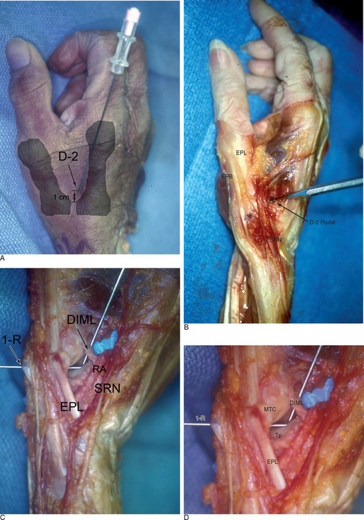
Access to medial osteophytes may sometimes be difficult. Hence, I have found the use of a distal/dorsal (D-2) accessory portal to be of some value. Its main utility is that it allows one to look down on the trapezium rather than across it, which facilitates resection of medial osteophytes. This accessory portal allows views of the dorsal capsule with rotation of the scope and facilitates triangulation of the instrumentation. It is situated in the dorsal aspect of the first web space. An anatomical study of five cadaver hands revealed that the D-2 portal surface landmark is ulnar to the EPL tendon and 1 cm distal to the V-shaped cleft at the juncture of the index and thumb metacarpal bases. The portal lies just distal to the dorsal intermetacarpal ligament (DIML).8
The DIML is an extracapsular ligament that originates from the dorsoradial aspect of the index metacarpal radial to the extensor carpi radialis longus insertion. It inserts onto the palmar-ulnar tubercle of the base of the thumb metacarpal along with the POL and UCL.4 A trocar placed through the D-2 portal was found to pass through the first dorsal interosseous muscle and penetrate the DIML, entering the joint either through or between the ulnar collateral ligament (UCL) and the posterior oblique ligament (POL) (Figure 2.1a through d). Branches of the superficial radial nerve passed within 3.2 mm (range 1 to 5 mm) of this portal. The radial artery was 3.8 mm away (range 3 to 5 mm) from this portal, the FDMA was within 2.8 mm (range 2 to 4 mm) of this portal, and the cephalic vein was within 2.8 mm (range 1 to 5 mm) of this portal. On average, the D-2 portal was 17.2 mm from the 1-U portal (range 12 to 20 mm).
< div class='tao-gold-member'>


Stay updated, free articles. Join our Telegram channel

Full access? Get Clinical Tree


