Transtibial Amputation
Introduction
Myofascial posterior skin flap initially used to treat peripheral vascular disease
Mixed data on necessity of tibia/fibula bone bridge
Ideal tibial bone length 2.5 cm for each 30 cm of height
Patient Selection
Ischemic limbs need thorough workup before amputation
Ankle-brachial index greater than 0.5
Transcutaneous oxygen saturation greater than 20 to 30 mm Hg
Albumin level greater than 2.5 g/dL
Absolute lymphocyte count greater than 1,500/μL
Of amputated limbs, 9% to 15% progress to further amputation
Indications
High-energy trauma, avascular limb, congenital deformity, tumor, infection, chronic pain
No clear absolute indications with validated outcomes exist for amputation in trauma setting
Loss of plantar sensation not an indication in the early trauma patient
Patients with dysvascular limbs considered for amputation have nonreconstructible injuries or are not revascularization candidates
When considering bone-bridge synostosis, ensure patient can safely endure longer tourniquet times (115 versus 71 minutes) and longer surgical times (179 versus 112 minutes)
Authors recommend reserving bone-bridge synostosis for young, healthy, active patients
Those with fibular instability and disruption of interosseous membrane may benefit from bone-bridge synostosis as primary or revision amputation
Contraindications
Dysvascular limbs not meeting preoperative assessment criteria
Consider procedures for optimization of wound healing or higher level amputation
Procedure
Room Setup/Patient Positioning
Supine position on standard table
Bump under ipsilateral limb
Thigh tourniquet
Special Instruments/Equipment/Implants
Basic major orthopaedic set
Oscillating saw
Drill
Amputation knife optional
Nonabsorbable suture ligatures for arteries; simple ligatures or vessel clips for veins
Suction drain
Small or large fragment set for bone-bridge synostosis or screw fixation
Appropriate bone-bridge fixation device
Chisel or osteotome
C-arm
| Video 96.1 Transtibial Amputation. COL James R. Ficke, MD; MAJ Daniel J. Stinner, MD (5 min) |
Surgical Technique

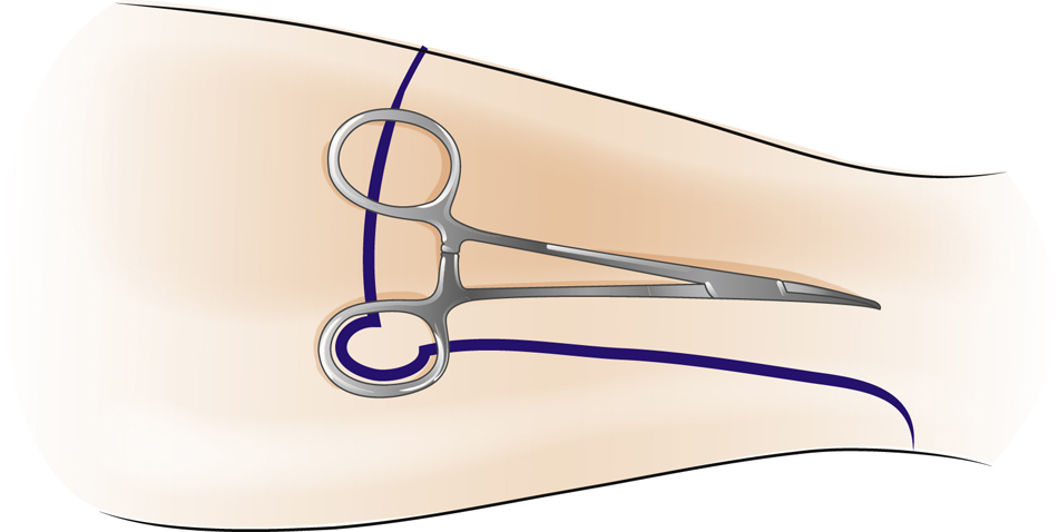
Figure 1Illustration depicts a method of minimizing redundant skin “dog ears.”

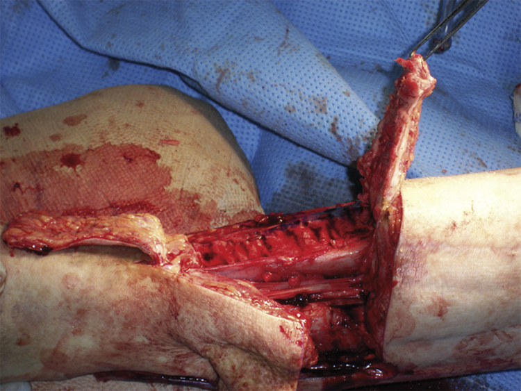
Figure 2Intraoperative photograph shows elevation of the periosteal flap. Multiple small bone fragments can be seen.

Figure 3Intraoperative photographs show transtibial amputation. A, Extended posterior flap with tapered gastrocnemius. Note that the tibial bevel does not encroach on the medullary canal. B, Myodesis is achieved with large braided suture to bone. The periosteal flap can be seen underneath, and the submuscular drain is in place. C, The completed myodesis is shown. The posterior skin flap overlap is traced before suprafascial skin excision.
Perform exsanguination
Mark tibial tubercle
Stay updated, free articles. Join our Telegram channel

Full access? Get Clinical Tree


